Summary
Here, we present the modifications necessary to a well characterized and commonly used small animal ferric chloride-induced (FeCl3) carotid artery injury model for use in a large animal vascular injury model. The resulting model can be utilized for pre-clinical trial assessment of both prophylactic and thrombolytic pharmacological and mechanical interventions.
Abstract
Occlusive arterial thrombosis leading to cerebral ischemic stroke and myocardial infarction contributes to ~13 million deaths every year globally. Here, we have translated a vascular injury model from a small animal into a large animal (canine), with slight modifications that can be used for pre-clinical screening of prophylactic and thrombolytic agents. In addition to the surgical methods, the modified protocol describes the step-by-step methods to assess carotid artery canalization by angiography, detailed instructions to process both the brain and carotid artery for histological analysis to verify carotid canalization and cerebral hemorrhage, and specific parameters to complete an assessment of downstream thromboembolic events by utilizing magnetic resonance imaging (MRI). In addition, specific procedural changes from the previously well-established small animal model necessary to translate into a large animal (canine) vascular injury are discussed.
Introduction
Stroke therapy is largely modeled after coronary artery disease treatment, mainly because interventions in cardiovascular disease have responded well to drug therapy and endovascular interventions1. These treatments, however, have not successfully translated to cerebral infarction. The difficulties with the current stroke treatment are that the recombinant tissue plasminogen activator (rTPA) cannot be reversed, and that administration carries a significant 6.4% risk of hemorrhagic conversion2,3,4. The resulting morbidity and mortality limits its use to a small, often unattainable window5. Also, restenosis and occlusion occur often after initial thrombolysis, reversing initial neurological improvement. In summary, there is a narrow temporal window to administer rTPA that excludes the large majority (~90%) of patients who suffer ischemic cerebrovascular insults.
The role of intravenous antiplatelet therapy has shown promise in treating ischemic stroke with improved vessel recanalization, survival and outcome2. Unfortunately, these drugs have a predictable side-effect of intra-cranial and extra-cranial hemorrhage, largely because there is no way to adequately reverse or control their activity2. While effective in preventing platelet aggregation, the risk of hemorrhage and the inability to reverse their activity have precluded their use in the routine care of stroke patients. A need, therefore, exists for potent antithrombotic drugs that act alone or in combinations to prevent and lyse clots yet have a safety profile that will allow the use in a closed, low volume space such as the brain, where hemorrhage is poorly tolerated.
Understanding the mechanism of arterial thrombosis and re-stenosis, and evaluating thrombolytics and drugs that prevent re-stenosis, requires both small and large animal models as a part of pre-clinical drug development. Ferric chloride-induced vascular injury is a widely utilized technique to rapidly and accurately induce the formation of thrombi in exposed blood vessels of mice, rats, guinea pigs, and rabbits6,7,8,9,10,11,12. These smaller species offer several advantages including ease of genetic manipulation, inexpensive animal purchase, and low per diem housing costs. Unfortunately, small animal experiments negate multiple blood draws during the surgery to access platelet reactivity, blood gas analysis, and inflammatory response. More importantly, large animals much more closely mimic human platelet physiology6,13. The FeCl3 carotid artery injury model has played a predominant role in the study of the pathophysiology of thrombosis, in the validation of novel anti-platelet and anti-coagulant drugs, and in the discovery of potential thrombolytics6,7,8,9,10,11,12. Previous models in mice, rats, guinea pigs, and rabbits have provided ease and flexibility for the genetic manipulation, but translatable pre-clinical models are critical to patient dosing and toxicity studies of potential therapeutics6,13. Although several models of thrombotic disorders have been developed in mice, large animal models of thrombosis that are applicable to the peripheral vascular disease, stroke and myocardial infarction are few and far. The first thrombosis models in monkeys, dogs, and pigs focused on stenosis, applying hemostats and later cylinders to vessels, commonly resulting in cyclic flow reductions14,15,16. Instead of an occlusive thrombus at the site of the endothelial damage as in the ferric chloride model, the thrombus in these models resulted in cyclic thrombosis, distal embolization and return to normal blood flow. In comparison, the ferric chloride model modified here in a large animal, results in an occlusive thrombus at the injury site and is stabilized and verified by angiography before thrombolytic treatment. Provided that the investigator has ample funds for per diem and purchase of canines and adequate surgical expertise, we detail here a large canine model of vascular injury to allow laboratories to study thrombosis utilizing surgical, imaging and histological techniques.
Subscription Required. Please recommend JoVE to your librarian.
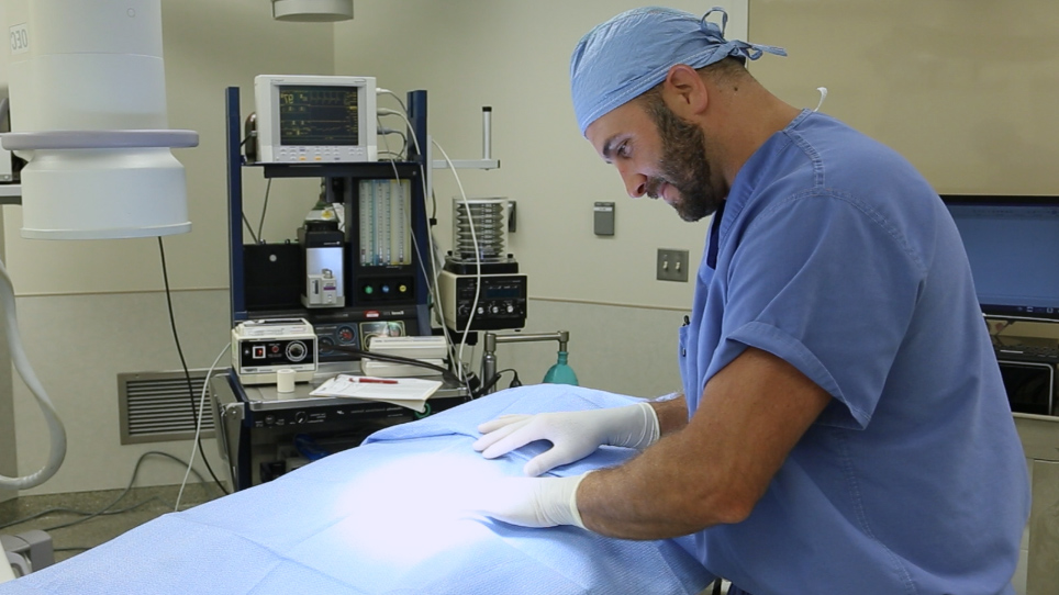
Protocol
The investigations described conform to the Guidelines for the Care and Use of Laboratory Animals of the National Institutes of Health and were approved by The Ohio State University Institutional Animal Care and Use Committee (#2015A00000029). All surgical manipulations were performed under deep anesthesia and the animals did not experience pain at any stage during the procedure. All experiments described were non-recovery.
1. Preparation
- Freshly prepare 50 mL of ferric chloride (FeCl3) at 50% (w/v) diluted in deionized water.
- Cut the umbilical tape into 5.5 cm pieces and soak in the freshly prepared 50% FeCl3 solution 1 week before the procedure.
NOTE: The umbilical tape used is thick and takes some time to absorb the 50% FeCl3 solution. - Fast each canine overnight before surgery.
- Label cryovials for plasma and urine collection, 50 mL sterile centrifuge tubes for all organ storage, and 10% formalin containers for brain and carotid fixation for H&E staining with animal identification, date of birth, date of surgery, and treatment.
- Prepare 100 mL of each solution for each canine procedure: 4% paraformaldehyde in phosphate buffered saline (pH 7.4), 10% neutral buffered formalin, and 2% 2,3,5-triphenyltetrazolium chloride [TTC in PBS (pH 7.4), store in the dark].
- Acquire ample liquid nitrogen for all cryovials and 50 mL tissue tubes for flash freezing for the day of the procedure.
- Acquire two ADP/collagen cartridges for PFA-100 (Platelet Function Analyzer) platelet reactivity for each canine for each time point of interest.
- Acquire one 4.5 mL sodium citrate blood collection tube, for each blood draw before sacrifice, for plasma separation and storage. At sacrifice, draw 16 blood collection tubes for the plasma storage. After each blood collection tube is full, spin at 4,000 x g for 30 s to isolate platelet rich plasma and transfer clear layer into cryovials. Flash freeze in liquid nitrogen before storage at -80 °C.
- Acquire one 4.5 mL lithium heparin blood collection tube, for each PFA-100 time point of interest to record platelet reactivity. At each blood draw, introduce 800 µL of whole blood from a lithium heparin blood collection tubes into each ADP/collagen cartridge without air bubbles to follow the platelet reactivity.
- Acquire two EDTA K3 blood collection tubes (1.8 mg of anhydrous EDTA per 1 mL of blood), for complete blood count (CBC) at baseline and at time of sacrifice.
NOTE: This volume is adequate for most core facilities to run each twice in case of mechanical error. Although a CBC was run at both the baseline before injury and at the time of sacrifice, investigators can add more blood draws as approved in their animal protocols for additional analyses. Check with the facility as to volumes and delivery requirements for their equipment. - Prepare a 2 mL heparinized syringe by filling and coating the syringe the day of the procedure for blood gas analysis at time of sacrifice.
NOTE: Check with the facility as to volumes and delivery requirements for their equipment.
2. Canine Carotid Artery Occlusion
- Weigh an adult (>18 months) beagle canine and pre-medicate with intramuscular acepromazine (0.025-.2 mg/kg) followed by an initial intraperitoneal injection of ketamine (5-20 mg/kg) and midazolam (0.01-.25 mg/kg). Induce deeper anesthesia with 1-5% isoflurane based on the heart rate, respirations, and jaw tone.
NOTE: Exact anesthesia is dictated by the weight and the approved animal protocol. Although injections may result in pain for the animals, the duration of this discomfort will be minimal (less than 3 s). - Prior to taking the animal to the surgery table, remove hair by clipping. Apply lubricating ophthalmic ointment to the anesthetized animal's eyes to prevent dryness.
- Place the animal in supine position using a positioning system and attach electrocardiography (ECG) leads to foot pads. Intubate through the trachea with a 6.5 mm cuffed endotracheal tube, mechanically ventilate at 14 breaths/min, and maintain each dog with a continuous inhalation of 1-5% isoflurane (depends on the approved animal protocol).
- To determine the extent of canalization of the carotid and the length of the occlusive thrombus, perform angiography before injury, before MRI, and at time of sacrifice (see below for additional instructions).
- Insert a femoral arterial sheath and a plastic catheter and secure with a loose 0-silk suture. Temporarily close the sheath and catheter during the transport and imaging studies. See canine angiography below for additional instructions).
- For blood draws, insert a 16G x 45 mm intravenous catheter through the intact skin distal to the femoral arterial sheath cut down site. Advance the catheter into the surgical exposure and visualize it as it enters into the exposed femoral vein immediately adjacent to the arterial sheath. Once fully inserted into the vein, withdraw the needle, cap the catheter with a 3-way stopcock, and flush it with 2 mL of 0.9% sterile saline. Secure the catheter to the skin with a 0-silk suture.
- Make an 8-10 cm incision of the skin directly on the top of the right common carotid artery region and dissect the fascia to isolate the vessel from the surrounding tissue.
- Carefully introduce forceps between the carotid artery and the vagus nerve to separate. Avoid touching the carotid artery more than necessary as it may cause constriction of the vessel.
- Dry the exposed area of the artery with the sterile gauze to avoid FeCl3 solution dilution.
- Place the Doppler flow probe around the carotid allowing amble space to wrap the umbilical tape around the carotid without touching the flow probe and start recording the blood velocity and continue throughout the procedure (Figure 1 Top). Record the baseline flow for ~ 5 min before applying the umbilical tape.
NOTE: Baseline flow before FeCl3 umbilical tape placement usually averages around 250 mL/min. - Wrap the prepared FeCl3 umbilical tape around the carotid using a hemostat immediately below the flow probe without touching it for 15 min, remove, and discard.
NOTE: Occlusion is designated as zero flow mL/min. - Stabilize the occlusive thrombus for 45 min. Add additional saline to the probe as needed to maintain a signal.
NOTE: Depending on the age, gender and the type of canine, additional placements of the prepared FeCl3 umbilical tape may be necessary for allowing 30 min for occlusion to occur before re-application. No differences were observed in thrombosis by ferric chloride application with gender in beagles. It was not found necessary with this model to reapply a fresh FeCl3 umbilical tape more than once and have never deviated from the FeCl3 umbilical tape preparation protocol. If the laboratory is using a different breed or age of canine, the FeCl3 umbilical tape should be applied to the initial subjects to make sure that the thrombosis protocol does not deviate, or a group of canines may be necessary to establish FeCl3 application time for that breed or age in their specific study. The occlusion times, in this experiment, range from 8-30 min after FeCl3 umbilical tape application. - To determine the stroke or hemorrhage volume in addition to the downstream thromboembolic events which may result from pharmacological interventions, transport canines with additional ketamine injection to a magnetic resonance imaging machine (see below for additional processing).
- While still under a deep surgical plane of anesthesia from a final angiogram/MRI (see below for imaging protocol), indicated by a lack of response to noxious stimuli, collect whole blood desired for later analysis, then open the chest by cutting through the ribs starting at the sternum, and excise the heart by cutting the blood vessels (exsanguination).
NOTE: In this experiment, whole blood (<10 mL) was drawn at baseline, 5 min after agent administration, and 60 min after infusion from the femoral sheath and saw no effect on the process of thrombolysis. At sacrifice, an unlimited volume can be drawn for additional plasma storage if approved in the animal protocol. Analyze, process and store all whole blood within 30 min of sacrifice. Ensure that blood is not drawn in excess of the approved percentage of the animal's body weight as detailed on the approved animal protocol. All experiments described were non-recovery. - Rinse the heart with 0.9% saline and flash freeze samples in the liquid nitrogen if desired.
- Before removing the carotids, measure the clot length by placing a ruler beside it and record. Place the tissue marker on the middle of the clot and at the same place on the contralateral carotid for ease in histology trimming. Mark the contralateral carotid at the same length.
NOTE: The average clot length with this model is 1.75 cm. - Remove the entire length of the clot on both vessels and fix in 10% formalin. Remove the contralateral carotid artery within the length marked by the tissue marker. Cut the contralateral carotid artery in half at the middle of the clot. Flash freeze half the contralateral carotid artery in liquid nitrogen for later analysis and fix the other half in 10% formalin to embed beside the injured carotid for histological analysis below.
- Remove organs for any desired analysis, rinse with 0.9% saline, and flash freeze in liquid nitrogen.
- Remove the skull with a circular bone saw. Slowly remove the skull without damaging the brain beneath.
- After the brain is removed from the skull, rinse it in cold 0.9% saline, and cut two 4 mm sections from the middle of the brain, cutting laterally with a sharp scalpel. Place one of the 4 mm sections into 2% TTC for ischemic demarcation (see additional processing for brain TTC), while the other slice placed in 10% formalin for 7 days for H&E staining (see additional processing for brain H&E).
3. Canine Angiography
NOTE: This is shown in Figure 2 and is done during the surgery at time points of interest.
- Feel for a pulse in the right inguinal region and make a 3-5 cm ventral sagittal incision.
- Expose the femoral artery and separate it from the surrounding tissues using blunt dissection.
- Use a right-angle hemostat to place a 0-silk suture around the distal end of the exposed artery.
- Tie the distal 0-silk suture and apply slight tension to the artery by clamping the loose 0-silk suture ends to the skin with hemostats.
- Place a second 0-silk suture around the proximal end of the exposed segment.
- Puncture the artery midway between the proximal and distal 0-silk suture ties using an 18G introducer needle to pass a guide wire into the artery.
- Load an assembled 6Fr sheath and dilator onto the guide wire, grasping the wire as it exits from the assembly.
- Advance the dilator and sheath over the guide wire. Make sure to maintain a firm grasp of the wire as it protrudes from the dilator while advancing the dilator sheath assembly.
- After full insertion, tie the proximal 0-silk suture around the sheath to maintain hemostasis at the arterial puncture site.
- Simultaneously remove the dilator and the guide wire.
- Open the one-way valve to verify the presence of pulsatile blood flow and flush with 3-5 mL of normal saline.
- Connect the access sheath one-way valve to a fluid filled extension line attached to a pressure transducer to monitor and record invasive blood pressure measurements.
- Pre-load a 0.35" guide wire into a 4Fr. angiographic catheter and connect to the contrast injection manifold set with a Tuohy y-adapter.
- Pull the tip of the guide wire just inside the distal tip of the catheter and flush the entire assembly with saline.
- Insert the angiographic catheter into the hemostatic valve of the sheath and then advance the guide wire about 5 cm beyond the distal catheter tip.
- Under fluoroscopic guidance and using a retrograde trans-aortic approach, slowly advance the catheter into the aortic arch.
- Inject 2-3 mL of contrast agent diluted 1:1 with normal saline to identify the take-off of the brachiocephalic trunk.
- Use the guide wire to direct the catheter into the brachiocephalic trunk, and then selectively into the right common carotid artery.
- Remove the guide wire and inject 2-3 mL of diluted contrast to verify that the catheter is placed in the carotid.
- Inject 3-4 mL of undiluted contrast while recording angiographic runs using both digital subtraction and standard angiographic techniques.
- Repeat steps 3.18-3.21 for the repeated angiography at additional time points.
4. Magnetic Resonance- Diffusion Weighted Imaging (DWI) and T2 weighted imaging (T2WI) of canine brain
NOTE: This is shown in Figure 3A-3B.
- Ensure that the dog is under deep anesthesia.
- Ensure that the dog's physiological parameters are monitored throughout the imaging including ECG and heart rate.
- Ensure that the dog is lying in the supine position with its head in the bilateral openings of the brain coil.
- Enable the 3 Tesla (3T) magnet.
- Perform T2 weighted imaging (T2WI), a basic pulse sequences in the MRI.
NOTE: The sequence uses the differences in the T2 relaxation time of tissues at different pathological conditions. - Perform localizer imaging to acquire pilot images of a dog brain before the anatomical imaging using T2-weighted gradient echo imaging sequence and determine the field of view (FOV), number of slices and slice thickness to completely cover the brain.
- Use the following T2-parameters: slice thickness = 3 mm, TR = 4,000 ms, TE = 75 ms, ETL = 7, acquisition matrix = 320 x 256, FA = 180ᵒ, FOV = 320 x 320 pixels, image resolution = 2.4615 pixels per mm.
- Perform diffusion weighted imaging (DWI) using echo planer DTI sequence 4 h after MCA occlusion and immediately before sacrifice.
- Use the following DWI parameters: b = 1,500 s/mm2, slice thickness = 3 mm, TR = 4,600 ms, ET = 86 ms, ETL = 55, acquisition matrix = 140 x 140, FA = 90ᵒ, FOV = 231x257, image resolution = 0.9333 pixels/mm (Table 1).
5. Hematoxylin and Eosin (H&E) staining of the canine brain
NOTE: This is shown in Figure 3D.
- After 7 days in 10% formalin, embed the 4 mm brain sections in paraffin.
- Trim the paraffin blocks until they are level with a microtome, removing any additional paraffin from the top of the brain tissue, then cut the brain tissue at 4 µm and place one brain section on each 2" x 3" inch slide.
NOTE: Before staining, place all solutions in separate containers so that slides can be moved from one solution to another without drying out. - De-paraffinize each brain slide and hydrate with tap water.
- Place each brain slide in Hematoxylin 560 for 8 min.
- Rinse each brain slide in tap water.
- Differentiate each brain slide with 1% Acid Alcohol for 1 s three times.
- Rinse each brain slide in tap water.
- Blue each brain slides with 1% ammonium hydroxide for 1 s.
- Rinse each brain slide in running tap water for 2 min.
- Dehydrate each brain slide in 70% Ethanol (EtOH) for 1 s twelve times.
- Counterstain each brain slide in eosin for 1 min.
- Dehydrate each brain slide in 95% EtOH for 1 s twelve times.
- Dehydrate each brain slide in 100% EtOH.
- Clear each brain slide in xylene and then apply a 2" x 3" inch coverslip on top of the brain slide, removing air bubbles with resinous mounting media.
NOTE: All solutions are reused except water and ethanol for multiple days of staining and multiple slides. All processing after brain sections are placed on slides are done in glass staining jars, but plastic staining containers are commercially available also. Replace any solution when it becomes discolored and keep them tightly covered.
6. Hematoxylin and Eosin (H&E) staining of canine carotids
NOTE: This is shown in Figure 1 (Left).
- After 24-72 h in 10% formalin, cut the injured carotid in half at the middle of the clot and embed ~ 1 cm of injured carotid and ~ 1 cm of the contralateral control in the same paraffin cassette.
NOTE: The blood clot inside the carotid is fragile. Wait until the vessel has been fixed in 10% formalin before cutting so that the clot is not disturbed. Embedding both the injured and control carotid arteries in the same paraffin block will allow cutting at the same time at the same place in the blood vessel. - Trim paraffin blocks until level with a microtome, removing any additional paraffin. Then cut sections at 4 µm on to 25 mm x 75 mm x 1 mm slides without rotating the paraffin block so that the contralateral carotid sections are placed at the top of the slide.
NOTE: Three carotid sections were placed on to each slide, but one can add depending upon stains of interest. - De-paraffinize each carotid slide and hydrate in tap water.
NOTE: Unlike the brain slide which should be processed individually, carotid slides can be processed in bulk by placing in jars which fit several slides at a time without touching one another. - Stain each carotid slide in hematoxylin for 8 min.
- Rinse each carotid slide in tap water.
- Differentiate each carotid slide with 1% Acid Alcohol for 1 s three times.
- Rinse each carotid slide in tap water.
- Blue each carotid slide with 1% ammonium hydroxide for 1 s.
- Rinse each carotid slide in running tap water for 2 min.
- Dehydrate each carotid slide in 70% EtOH for 1 s twelve times.
- Counterstain each carotid slide in eosin or 1 min.
- Dehydrate each carotid slide in 95% EtOH for 1 s x 12.
- Dehydrate each carotid slide in 100% EtOH.
- Clear each carotid slide in xylene and add a 24 mm x 50 mm coverslip with resinous mounting media to remove the air bubbles.
7. 2,3,5-triphenyl-2H-tetrazolium Chloride (TTC) Staining of the Canine Brain
NOTE: This is shown in Figure 3C.
- Place the other 4 mm section removed from the middle of the brain immediately proximal to the H&E section into previously prepared 2% TTC (carotid artery occlusion). Incubate in the dark at 37 °C for at least 20 min, turning over the brain section for even staining every 5 min.
NOTE: After 20 min, the section should be cherry red on both sides. Additional time may be needed based on TTC freshness and thickness of tissue section. - Remove the TTC solution and replace it with 4% paraformaldehyde in PBS, pH 7.4 to optimize the color contrast.
- When the contrast between white and red staining is optimal, place TTC brain slices between clear plastic sheets, dry of excess fluid, and scan at high resolution for tracing of ischemic regions.
NOTE: Sections can be stored indefinitely in paraformaldehyde solution, but staining will fade.
Subscription Required. Please recommend JoVE to your librarian.
Representative Results
Following the detailed procedures herein will result in the development of a model that can be used for prophylactic or thrombolytic assessment of occlusive arterial interventions. Figure 1A shows baseline flow velocity and the resulting blood flow velocity before, during, and after treatment recorded by a commercial software. Data from this recording can be used to determine the percent of re-perfusion with carotid artery injury and treatment in this canine model. Figure 1B provides an example of both the contralateral (top) and injured (bottom) canine carotid sections stained with H&E that verifies the re-canalization status at the time of sacrifice. A multitude of software programs are available to analyze the perfused area of the vessel by tracing the blood vessel (without thrombus) which can be divided by total area of the vessel to arrive at a percent of canalization with each treatment. Figure 2 shows several examples of carotid artery angiography detailed in this canine imaging protocol which can be used to determine if the thrombus is occlusive. In addition, using the square flow probe as a marker, investigators can determine the length of the thrombus at each time point the angiogram is taken. Although, here we have presented images before injury, 60 min after vehicle treatment, and at the time of sacrifice, the investigator can tailor imaging to their needs. Figure 3 is the result of the magnetic imaging parameters applied to the canine brain ~ 4 h after carotid artery occlusion immediately before sacrifice, utilizing both diffusion weighted (Figure 3A) and T2 weighted imaging (Figure 3B) detailed in section 4 of this protocol. Although we only show one photo of the entire array taken at different levels in the brain, the size of both hemorrhage and stroke volume can be identified in different levels and areas of the brain and quantitated at each time point desired by the investigator. Quantitation is completed using the specific MRI machine software specific to the investigator's imager. The TTC staining results as shown in Figure 3C can be used to delineate brain tissue in which the cells are still metabolically active in addition to those that are not. TTC will be enzymatically reduced to result in red stained live cells whereas dead cells will not retain the TTC and thus will not be red. Lastly, the hematoxylin and eosin staining technique demonstrated in canine brain in Figure 3D will result in staining red blood cells bright red which can be used to verify areas where hemorrhage has occurred.

Figure 1: Monitoring of carotid blood flow. (A) Representative carotid artery blood velocity (mL/min) recorded from the Doppler probe from baseline before injury through sacrifice. (B) Representative H&E staining of both occluded carotid artery (bottom) and contralateral control (top). Magnification is at 20X. Please click here to view a larger version of this figure.

Figure 2: Monitoring of the carotid blood flow by angiography. Representative angiography view of right canine carotid artery. Images were taken at baseline before vehicle infusion (A), 60 min after vehicle infusion (B), and at time of sacrifice, 4.5 h after occlusion (C). Red arrows indicate the location on the vessel of the FeCl3-induced injury where the umbilical tape was placed. Please click here to view a larger version of this figure.

Figure 3: Representative images of canine brain after vehicle treatment. Magnetic Resonance Imaging (MRI) performed ~4 h after occlusion, immediately before sacrifice, utilizing diffusion weighted imaging (DWI, (A) and T2 weighted imaging (B). TTC staining of medial section at the time of sacrifice to delineate live from dead tissue (C). H&E staining of medial section, immediately proximal to TTC section, at time of sacrifice to delineate tissue which are metabolically active (red) from those that are not TTC is reduced to a red product when taken up by live cells and therefore, will result in sections that can be quantified with a multitude of software to trace live vs dead regions. (D). Please click here to view a larger version of this figure.
| Experimental Parameter | T2-weighted | Diffusion weighted |
| Repetition time (TR) ms | 4000 | 4600 |
| Echo time (TS) ms | 75 | 86 |
| Flip angle, degrees | 180 | 90 |
| Acquisition matrix | 320 x 256 | 231 x 257 |
| Number of averages | 2 | 4 |
| In plane image resolution (pixels/mm) | 2.4615 | 0.9333 |
Table 1: Magnetic Resonance Imaging (MRI) parameters. MRI parameters that were developed for canine T2- and Diffusion-weighted images to maximize for assessment of stroke and hemorrhage volume measurement in canine carotid artery thrombosis model.
Subscription Required. Please recommend JoVE to your librarian.
Discussion
The FeCl3 induced vascular injury model is widely used to study thrombosis in small animals and is easy to translate into a large animal, pre-clinical model with a multitude of advantages. Slight modifications to adapt the protocol into a canine allow the utilization of both magnetic resonance imaging to assess stroke and hemorrhage volumes after a pharmacological intervention and angiography to assess vessel canalization before, during, and after treatment. Other thrombotic large animal models have not studied stabilized occlusive thrombi at the site of injury and therefore cannot utilize angiography and histology of the carotid artery to assess the extent of re-canalization for each prophylactic or thrombolytic treatment. In addition to the advantages of a large animal which include ample tissue for analysis (plasma, urine, organs for toxicity studies, etc.), the large animal model much more closely mimics the heart rate, blood pressure, and coagulation cascade in humans than rodent models. Adequate size of both the brain and carotid arteries, result in histological and biological material that can be used for a plethora of inflammatory and biochemical investigations on each experimental animal. Another advantage of modifying the FeCl3-induced vascular model into a canine is that a much larger blood volume can be extracted during the experiment without modifying platelet reactivity or thrombus formation such that blood gas and complete blood counts can be monitored throughout the injury and drug infusion. In addition, the ferric chloride injury has been attributed to the oxidant damage; therefore, it will mimic atherosclerotic damage that precedes clinical stroke and myocardial infarction much more closely than the other types of published arterial injury models7. Lastly, mechanical interventions such as thrombectomy, which are routinely used clinically, can follow pharmacological treatment so that re-stenosis and the status of the endothelial wall health can be addressed with ample histological tissue for multiple investigations on the same canine.
Limitations of this model are few but need to be considered. First, although the time point, and treatment chosen in this publication (sacrifice 4.5 h after carotid artery injury, vehicle) did not result in a measurable stroke or hemorrhage, this method is clinically relevant for the investigation of novel anti-thrombotic and thrombolytic agents to determine efficacy and dosing. Indeed, both stroke and/or hemorrhage with the initial thrombotic insult or with pharmacological treatment (i.e., rTPA) do occur with this model. Secondly, canine costs for purchasing, shipping, genetic manipulation, and per diem are quite high and cost prohibitive until a drug is well-characterized in rodent models.
Critical steps in the protocol center on platelet activation, aggregation and adhesion, the crux of thrombosis. Since the Platelet Function Analyzer (PFA-100) can be performed with 1600 µL of whole blood in duplicate, this method is simple and easy to track platelet reactivity throughout the procedure without affecting blood homeostasis. Further ex vivo studies into platelet activity using impedance or lumi-aggregometry can be performed before vascular injury without affecting experimental thrombosis as long as the last blood draw is >1 week before surgery in addition to after injury and treatment at time of sacrifice. As previously discussed, additional applications of umbilical tape soaked in 50% FeCl3 may be necessary depending on gender, breed, or age of each canine. We allowed 30 minutes after injury for occlusion and re-applied fresh 50% FeCl3 for another 15 min if necessary. This process did not result in a significant difference in thrombolytic deviation with age or gender using adult beagles. In this study, we have elucidated the critical and state-of-the-art diffusion weighted imaging (DWI), a MR imaging based on measurement of Brownian motion (random diffusion of particles) of water molecules within the tissue voxel. This technique is useful in detection of acute ischemic stroke among other pathologies such as tumors by showing diffusion in hyper-cellular tissues or those with cellular swelling is low with higher diffusion coefficient17,18,19. Diffusion maps vary with the diffusion of water molecules in the brain tissue17,18,19. The B-value measures the gradient for diffusion of H2O molecules. In the ischemic injury site the free water experiences strongest signal attenuation at higher B-values19.
In addition to use of this canine protocol for studies in dosing, toxicity, and efficacy of prophylactic and thrombolytic agents, the size of a canine makes experiments in mechanical thrombectomy clinically relevant and easily achievable. Carotids can be easily processed, stained, and assessed utilizing protocols for human histology for study of immune cell infiltration after clot retrieval. These studies will be our next detailed protocols.
Subscription Required. Please recommend JoVE to your librarian.
Disclosures
None
Acknowledgments
We would like to thank the Center for Cognitive and Behavioral Brain Imaging at The Ohio State University for their financial and scientific support to develop and perform canine magnetic resonance imaging.
Materials
| Name | Company | Catalog Number | Comments |
| 1/8” umbilical tape | Jorgensen Laboratories Inc., | #J0025UA | for ferric chloride application |
| 4% paraformaldehyde in PBS | Alfa Aesar | AAJ61899AP | |
| 10% neutral buffered formalin | Richard-Allan Scientific | 5701 | |
| 2% 2,3,5-triphenyltetrazolium chloride (TTC in PBS, pH 7.4) | Sigma Aldrich | T8877 | |
| ADP/Collagen cartridges | Siemens Diagnostics | B417021A | |
| 4.5 ml 3.2% sodium citrate blood vacutainer | Becton Dickinson | BD 369714 | |
| 4.5 ml lithium heparin vacutainer | Becton Dickinson | BD 368056 | |
| EDTA K3 vacutainers | Becton Dickinson | BD455036 | |
| Doppler flow probe | Transonic Systems Inc | MA2.5PSL | |
| Hematoxylin 560 | Surgipath | 3801570 | |
| Eosin | Surgipath | 3801602 | |
| LabChart Software | ADInstruments Inc. | ||
| Prisma Fit 3 tesla (3T) magnet | Siemen's Diagnostics | ||
| Sodium heparin for injection (to coat blood gas syringe) | NovaPlus | 402525D | |
| HUG-U-VAC positioning system | DRE Veterinary | 1320 |
References
- Adams, H. P. Jr Stroke: a vascular pathology with inadequate management. Journal of Hypertension Supplement. 21 (5), S3-S7 (2003).
- Lansberg, M. G., Bluhmki, E., Thijs, V. N. Efficacy and safety of tissue plasminogen activator 3 to 4.5 hours after acute ischemic stroke: a metaanalysis. Stroke. 40 (7), 2438-2441 (2009).
- Nagel, S., et al. Therapy of acute basilar artery occlusion: intraarterial thrombolysis alone vs bridging therapy. Stroke. 40 (1), 140-146 (2009).
- Ciccone, A., Motto, C., Abraha, I., Cozzolino, F., Santilli, I. Glycoprotein IIb-IIIa inhibitors for acute ischaemic stroke. The Cochrane database of systematic reviews. 3 (3), (2014).
- The National Institute of Neurological Disorders and Stroke rt-PA Stroke Study Group. Tissue Plasminogen Activator for Acute Ischemic Stroke. New England Journal of Medicine. 333 (24), 1581-1588 (1995).
- Leadley, R., Chia, L., Rebellob, S., Gagnon, A. Contribution of in vivo models of thrombosis to the discovery and development of novel antithrombotic agents. Journal of Pharmacological and Toxicological Methods. 43 (2), 101-116 (2000).
- Bodary, P. F., Eitzman, D. T. Animal Models of Thrombosis. Current Opinion In Hematology. 16 (5), 342-346 (2009).
- Sachs, U. J. H., Nieswandt, B. In vivo thrombus formation in murine models. Circulation Research. 100 (7), 979-991 (2007).
- Bonnard, T., Hagemeyer, C. E. Ferric Chloride-induced Thrombosis Mouse Model on Carotid Artery and Mesentery Vessel. Journal of Visualized Experiments. (100), e52838 (2015).
- Kurz, K. D., Main, B. W., Sandusky, G. E. Rat model of arterial thrombosis induced by ferric chloride. Thrombosis Research. 60 (4), 269-280 (1990).
- Karatas, H., Erdener, S. E., et al. Thrombotic distal middle cerebral artery occlusion produced by topical FeCl(3) application: a novel model suitable for intravital microscopy and thrombolysis studies. Journal of Cerebral Blood Flow and Metabolism. 31 (6), 1452-1460 (2011).
- Li, W., McIntyre, T. M., Silverstein, R. L. Ferric chloride-induced murine carotid arterial injury: A model of redox pathology. Redox Biology. 1 (1), 50-55 (2013).
- Vilahur, G., Padro, T., Badimon, L. Atherosclerosis and Thrombosis: Insights from Large Animal Models. Journal of Biomedicine and Biotechnology. 2011, 1-12 (2011).
- Coller, B. S., Folts, J. D., Smith, S. R., Scudder, L. E., Jordan, R. Abolition of in vivo Platelet Thrombus Formation in Primates with Monoclonal Antibodies to the Platelet GPIIb/IIIa Receptor. Correlation with Bleeding Time, Platelet Aggregation, and Blockade of GPIIb/IIIa Receptors. Circulation. 80 (6), 1766-1774 (1989).
- Folts, J. An in vivo Model of Experimental Arterial Stenosis, Intimal Damage, and Periodic Thrombosis. Circulation. 83 (6 Suppl), (1991).
- Yasuda, T., et al. A canine model of coronary artery thrombosis with superimposed high grade stenosis for the investigation of rethrombosis after thrombolysis. Journal of the American College of Cardiology. 13 (6), 1409-1414 (1989).
- Schob, S., et al. Correlation Between Aquaporin 4 Expression and Different DWI Parameters in Grade I Meningioma. Molecular Imaging and Biology : MIB : the Official Publication of the Academy of Molecular Imaging. 19 (1), 138-142 (2017).
- Schob, S., et al. Diffusion-Weighted Imaging Using a Readout-Segmented, Multishot EPI Sequence at 3 T Distinguishes between Morphologically Differentiated and Undifferentiated Subtypes of Thyroid Carcinoma-A Preliminary Study. Translational Oncology. 9 (5), 403-410 (2016).
- Schob, S., et al. Diffusion-Weighted MRI Reflects Proliferative Activity in Primary CNS Lymphoma. Public Library of Science One. 11 (8), e0161386 (2016).