This report describes techniques to isolate and purify sulfated glycosaminoglycans (GAGs) from biological samples and a polyacrylamide gel electrophoresis approach to approximate their size. GAGs contribute to tissue structure and influence signaling processes via electrostatic interaction with proteins. GAG polymer length contributes to their binding affinity for cognate ligands.
Method Article
Detection of Glycosaminoglycans by Polyacrylamide Gel Electrophoresis and Silver Staining
In This Article
Summary
Abstract
Sulfated glycosaminoglycans (GAGs) such as heparan sulfate (HS) and chondroitin sulfate (CS) are ubiquitous in living organisms and play a critical role in a variety of basic biological structures and processes. As polymers, GAGs exist as a polydisperse mixture containing polysaccharide chains that can range from 4000 Da to well over 40,000 Da. Within these chains exists domains of sulfation, conferring a pattern of negative charge that facilitates interaction with positively charged residues of cognate protein ligands. Sulfated domains of GAGs must be of sufficient length to allow for these electrostatic interactions. To understand the function of GAGs in biological tissues, the investigator must be able to isolate, purify, and measure the size of GAGs. This report describes a practical and versatile polyacrylamide gel electrophoresis-based technique that can be leveraged to resolve relatively small differences in size between GAGs isolated from a variety of biological tissue types.
Introduction
Glycosaminoglycans (GAGs) are a diverse family of linear polysaccharides that are a ubiquitous element in living organisms and contribute to many basic physiological processes1. GAGs such as heparan sulfate (HS) and chondroitin sulfate (CS) may be sulfated at distinct positions along the polysaccharide chain, imparting geographic domains of negative charge. These GAGs, when tethered to cell-surface proteins known as proteoglycans, project into the extracellular space and bind to cognate ligands, allowing for the regulation of both cis- (ligand attached to the same cell) and trans- (ligand attached to neighboring cell) signaling processes2. Furthermore, GAGs also perform critical roles as structural elements in tissues such as the glomerular basement membrane3, the vascular endothelial glycocalyx4 and pulmonary epithelial glycocalyx5, and in connective tissues such cartilage6.
The length of GAG polysaccharide chains varies substantially according to its biological context and can be dynamically lengthened, cleaved, and modified by a highly complex enzymatic regulatory system7. Importantly, the length of GAG polymer chains contributes substantially to their binding affinity for ligands and, subsequently, to their biological function8,9. For this reason, determination of the function of an endogenous GAG requires appreciation of its size. Unfortunately, unlike proteins and nucleic acids, very few readily available techniques exist to detect and measure GAGs, which has historically resulted in relatively limited investigation into the biological roles of this diverse polysaccharide family.
This article describes how to isolate and purify GAGs from most biological tissues, and, also, describes how to use polyacrylamide gel electrophoresis (PAGE) to evaluate the length of the isolated polymers with a fair degree of specificity. In contrast to other, highly complex (and often mass spectrometry-based) glycomic approaches, this method can be employed using standard laboratory equipment and techniques. This practical approach may, therefore, expand investigators' ability to determine the biological role of native GAGs and their interaction with contextually important ligands.
Protocol
All biological samples analyzed in this protocol were obtained from mice, under protocols approved by the University of Colorado Institutional Animal Care and Use Committee.
1. Heparan sulfate isolation
- Delipidation of tissue samples
NOTE: Delipidation is an optional step for fat-rich tissues.- Make a 1:1 mixture of methanol and dichloromethane. Prepare approximately 500 μL per sample; larger pieces of tissue may require up to 1 mL.
- Place each tissue sample into a small glass container with a lid for delipidation.
NOTE: Sample mass may vary per tissue of interest and experimental needs. 50 mg or less is typically sufficient for adequate GAG yield, but some optimization may be required by the end-user. - Add 500 μL of the methanol:dichloromethane solution to each glass container and mix. Ensure all solid tissue samples are completely submerged in solvent.
NOTE: Use serological pipettes or plastic conical tubes to mix and handle the methanol/dichloromethane solution; other plastics may dissolve. - Place samples on a shaker (in secured rack) in a chemical fume hood and agitate gently for 1 h.
- Agitate the samples to mix and centrifuge at 17,000 x g for 5 min at 4 °C.
- Carefully pipette out the organic solvent fraction (supernatant). This is the lipid fraction - it does not contain GAGs and can be discarded.
NOTE: Try not to disturb the tissue as small pieces could be lost. It is preferable to leave some of the organic layer in the sample container, rather than risking the sample loss. - Leave the lid of the glass container open and let it evaporate overnight in the hood. Change the tube for the next step as those tubes will not survive further processing.
- Once the solvent mixture has fully evaporated, proceed to mechanical disintegration (if residual sample is >50 mg) or Actinase E digestion (< 50 mg).
- Mechanical disintegration of solid tissue (optional; for larger tissue samples)
NOTE: Most smaller pieces of solid tissue (approximately 50 mg or less) should dissolve completely during the digestion step. However, larger samples will require mechanical disintegration.- Flash-freeze samples of interest by placing them in an appropriately sized polypropylene tube and placing the closed tube into liquid nitrogen. Allow the sample to freeze until completely solid.
- Using a clean mortar and pestle, grind frozen samples into a powder-like consistency.
- Proceed directly to Actinase E digestion (Step 1.4).
- Sample desalting and concentration
NOTE: This is an optional step and only required for dilute liquid tissue samples (e.g., broncho-alveolar lavage fluid (BALF) or plasma).- Pool liquid samples into appropriate experimental groups (e.g., by biological replicate, or experimental group)
- Place total sample volume into a 500 μL centrifugal filter column with a molecular weight cut-off (MWCO) of 3,000 Da.
- Spin for 30 min at 14,000 x g at room temperature. Repeat as needed if the desired sample volume exceeds capacity of the centrifugal filter column.
- Wash each column 3x with 400 μL of deionized, filtered water. Discard the flow through.
- Invert the filter and spin for 1 min at 2,000 x g in fresh, appropriately labeled collection tubes. Freeze at -80 °C or proceed to next step.
- Actinase E digestion
NOTE: This step is required to digest and ultimately remove protein contaminants from your sample.- Mix samples 1:1 with recombinant Actinase E to a desired concentration of 10 mg/mL. Add appropriate volume of 10x digestion buffer concentrate. Desired final concentration: 0.005 M calcium acetate and 0.01 M sodium acetate, pH 7.5. For example: 190 μL of liquid sample, 190 μL of 20 mg/mL Actinase E, 20 μL of 0.05 M calcium acetate, 0.1 M sodium acetate.
- Agitate samples gently to mix, then digest for 48-72 h at 55 °C (up to 7 days for whole tissue).
- Heat to 80 °C for 15-20 min to heat inactivate Actinase E.
- Freeze the sample at -80 °C or continue to step 1.5.
- Sample desalting and concentration
NOTE: If a low mass of the GAG of interest is anticipated in the biological sample, or if conservation of consumable reagents (i.e., centrifugal filter columns) is desirable, samples can be pooled to produce a single concentrate per experimental group (e.g., by biological replicate, or experimental group).- Place total sample volume into a 500 μL centrifugal filter column with a MWCO of 3,000 Da.
- Spin for 30 min at 14,000 x g at room temperature. Repeat as needed, if the desired sample volume exceeds capacity of the centrifugal filter column.
- Wash each column 3x with 400 μL deionized, filtered water. Discard the flow through.
- Invert filter and spin for 1 min at 2,000 x g in fresh, appropriately labeled collection tubes (generally included with the centrifugal filter columns). Freeze at -80 °C or proceed to next step.
- Desiccation
- Either place the dissolved samples from step 1.5.5 in a rotational vacuum concentrator overnight or lyophilize the samples as detailed below.
- Freeze samples thoroughly either overnight in -80 °C or by dipping in liquid nitrogen.
- Pierce sample lids with an 18 G needle and place in the lyophilizer chamber. Add paper towels for packing as needed.
- Fix lyophilizer chamber to lyophilizer and freeze dry overnight (at least -40 °C, 0.135 Torr)
- Cation exchange column
- Resuspend desiccated samples in up to 400 μL of 8 M urea, 2% CHAPS solution (or, if pooling samples, resuspend to a max of (400/n) μL, where n = the number of samples in desired pool. Use as little of the detergent solution as possible.
- Equilibrate the cation exchange (IEX) column with 400 μL of 8 M urea, 2% CHAPS solution. Spin for 5 min at 2,000 x g at room temperature.
- Load 400 μL of sample/pooled samples into the IEX column. Spin for 5 min at 2,000 x g.
- Wash 3x with 400 μL of 8 M urea, 2% CHAPS solution. Spin for 5 min at 2,000 x g everytime.
- Elute 3x with 400 μL of 0.2 M NaCl. Spin for 5 min at 2,000 x g each. This is the low-affinity fraction - this can be retained for quality control purposes if desired.
- Elute 3x with 400 μL of 2.7 M (16%) NaCl. Spin for 5 min at 2,000 x g each. This fraction will contain the isolated glycosaminoglycans of interest - keep all of it!
- To desalt each eluted fraction, add methanol up to 80 vol% and incubate at 4 °C overnight. Spin each sample for 5 min at 2,000 x g. Recover the solid residue as this is dried de-salted glycosaminoglycan.
NOTE: Alternatively, skip this step and proceed to step 1.8 to de-salt the eluate without methanol.
- Sample desalting and concentration
- If necessary, pool eluted fractions (from 1.7.6) into appropriate experimental groups (e.g., by biological replicate, or experimental group).
- Place total sample volume into a 500 μL centrifugal filter column with a MWCO of 3,000 Da.
- Spin for 30 min at 14,000 x g at room temperature. Repeat as needed, if the desired sample volume exceeds capacity of the centrifugal filter column.
- Wash each column 3x with 400 μL deionized, filtered water. Discard flow through. Proceed directly to step 1.9.
- Chondroitin digestion
NOTE: The purpose of this step is to remove GAGs not of interest to the end user. In this case, chondroitinase is used to remove chondroitin. In the tissues used for generating Representative Results (broncho-alveolar lavage fluid and whole lung), digesting chondroitin sulfate leaves heparan sulfate as the primary residual GAG. End users may need to add additional digestion steps depending on their experimental aims.- Load 350 µL of digestion buffer (50 mM ammonium acetate with 2 mM calcium chloride adjusted to pH 7.0) to the centrifugal filter column without touching the membrane.
- Add 5 µL of recombinant chondroitinase ABC.
- Place the samples tube into a 37 °C oven and incubate for 1 h.
- Turn the column over and place into appropriately labeled collection tubes. Spin for 1 min at 2000 x g.
- Heat samples to 80 °C for 15-20 min to inactivate chondroitinase ABC.
- Sample desalting and concentration
- If necessary, pool chondroitin digested samples from step 1.9.5 into appropriate experimental groups (e.g., by biological replicate, or experimental group)
- Place the total sample volume into a 500 μL centrifugal filter column with a MWCO of 3,000 Da.
- Spin for 30 min at 14,000 x g at room temperature. Repeat as needed if desired sample volume exceeds capacity of the centrifugal filter column.
- Wash each column 3x with 400 μL deionized, filtered water. Discard the flow through.
- Invert filter and spin for 1 min at 2,000 x g in fresh, appropriately labeled collection tubes. Freeze at -80 °C or proceed to next step.
- Desiccation
NOTE: In this step, desiccation is necessary so that samples can be resuspended in the smallest possible quantity of water and running buffer.- Either place the chondroitin digested samples from step 1.10.5 directly in a rotational vacuum concentrator overnight or lyophilize as follows:
- Freeze samples thoroughly either overnight in -80 °C or by dipping in liquid nitrogen.
- Pierce sample lids with an 18 G needle and place in the lyophilizer chamber. Add paper towels for packing as needed.
- Fix lyophilizer chamber to lyophilizer and freeze dry overnight (at least 40 °C, 0.135 Torr)
2. Polyacrylamide gel electrophoresis of isolated and purified glycosaminoglycans
- Prepare solutions necessary for polyacrylamide gel electrophoresis (PAGE) in advance (Table 1).
NOTE: Select the percent acrylamide of resolving gel solution depending on the size of the glycosaminoglycans expected to be in the sample. 15% is recommended for resolving larger fragments (greater than 30 disaccharide subunits in length); 22% for smaller fragments (<20 disaccharide subunits in length). - Place empty cassette into the PAGE tank. Cast the resolving gel as follows: In 15 mL tube, mix 10 mL of resolving gel solution, 60 μL of 10% ammonium persulfate (must be freshly prepared), and 10 μL of TEMED (add TEMED last). Invert the tube gently 2-3x. Use pipette to quickly add the above 10 mL solution to cassette. Overlay with 2 mL of deionized, filtered water and allow the resolving gel to polymerize for 30 min.
NOTE: The following PAGE protocol has been optimized for a vertical PAGE system using 13.3 x 8.7cm (width x length) 1.0mm thick casting cassettes with a total volume of approximately 12mL. Other cassette systems can be used but may require optimization by the end-user. - After the resolving gel has fully polymerized, discard the overlaid water and cast the stacking gel as follows: in a 15 mL tube, mix 3 mL of the stacking gel solution, 90 μL of 10% ammonium persulfate (must be freshly prepared), 3 μL of TEMED (add TEMED last).
- Invert the tube gently 2-3x. Use a pipette to quickly add the stacking gel solution over the solidified resolving gel; fill cassette to brim. Fully insert comb included with the set up. Allow the stacking gel to polymerize for 30 min.
- Once the gel has polymerized, ensure the tape strip is removed from the bottom of the cassette, and place the cassette back into the PAGE tank assembly.
- Fill the upper and lower chambers with upper and lower chamber buffer, respectively.
- Dissolve the dried samples from step 1.11.4 in the minimum necessary volume of deionized, filtered water (at most, 50% of the volume of the wells in the PAGE gel). Mix 1:1 with sample loading buffer. Load the samples and the HS oligosaccharide "ladders" (see Table 1) into the gel.
- Pre-run the gel for 5 min at 100 V. Then run the gel at 200 V for 20-25 min (for a 15% polyacrylamide resolving gel), 40-50 min (for an 18% polyacrylamide resolving gel), 90-100 min (for 22% polyacrylamide resolving gel).
NOTE: Some optimization of the 200 V run time may be necessary. Phenol red migrates ahead of heparin oligosaccharides that are 2 polymer subunits in length (i.e., degree of polymerization 2, or dp2); bromophenol blue migrates ahead of dp10-dp14. Best results are obtained when the voltage is applied such that the phenol red band migrates almost, but not quite, to the bottom of the gel. Adjust run time accordingly.
3. Silver staining protocol
- Prepare all solutions necessary for silver staining in advance (Table 2).
NOTE: Do not directly touch the PAGE gel until it has been stained, developed, and placed in stop solution. Instead, manipulate the gel using clean plastic or glass tools. Directly handling the gel will result in finger-print distortions and other visible artifacts on the gel after staining. - Once the run is completed, disassemble cassette and extract gel in a clean, medium-large container filled with deionized, filtered water.
NOTE: To avoid directly handling the gel, use pipette tip or other plastic object to gently peel the gel away from the cassette while submerged in water. Gel may be fragile - handle carefully.- Discard the water. Stain the gel in Alcian blue staining solution for 5 min.
- Discard Alcian blue stain. Quickly rinse/wash 2-3x with deionized, filtered water until most of the Alcian blue staining solution has been removed.
- Allow to de-stain in deionized, filtered water overnight on rocker. Ensure that there is ample volume of deionized, filtered water to ensure any residual stain is fully washed off the gel overnight.
- Wash gel in 50% methanol (40 min total, change solution 2-3x).
- Wash gel in deionized, filtered water for 30 min. Discard water and repeat 3 more time for a total of 2 h, replacing the water each time.
- In a fresh, clean container, stain the gel for 30 min in silver nitrate staining solution.
- Quickly rinse/wash 2-3x in deionized, filtered water to fully remove the silver staining solution.
- Wash for 30 min in deionized, filtered water. Discard water and repeat 2x for a total of 90 min, replacing the water bath each time.
- Discard water and add developing solution.
- Once developing solution is added, carefully observe the gel and watch for the appearance of bands. Depending on the quality of the stain and the mass of the sample loaded, development can take anywhere from a few seconds to several minutes.
- As soon as the desired bands are visible, immediately discard developing solution and wash briefly with stop solution.
- Discard the stop solution wash and replace with fresh stop solution. Allow to soak for 1 h on a rocker or shaker.
- Wash in deionized, filtered water overnight (however, the gel can be imaged immediately after stop solution wash).
Results
Alcian blue is used to stain sulfated GAGs 10; this signal is amplified by use of a subsequent silver stain 11. Figure 1 provides a visual demonstration of the silver staining development process. As demonstrated, the Alcian blue signal representing GAGs separated by electrophoresis is amplified as the developing agent penetrates the polyacrylamide gel. Typically, the developing process will reduce silver and Alcian blue-stained GAGs in a density dependent fashion, with the edges of each band reducing first while the more densely staining regions in the center will stain last.
In the literature, the reported limit of detection of GAGs using PAGE-based approaches range from 0.5-1 µg12,13. To determine the limit of detection using our approach, heparan sulfate oligosaccharides of different polymer lengths (unfractionated heparin, dp20, dp10, dp6) were loaded onto a 22% polyacrylamide gel, then ran and stained as described above. Each oligosaccharide was loaded twice at two different masses: 1.0 µg and 0.5 µg. Figure 2 demonstrates that our technique can quite readily detect 0.5 µg of purified GAG. Notably, unfractionated heparin was least readily detected of the four different oligosaccharides tested, likely due to a wider distribution of polymer sizes that correspondingly reduces the density of each individual band.
To assess the efficiency of GAG purification from liquid biological samples, GAGs were isolated from two bronchoalveolar lavage (BAL) samples. As shown in Figure 3, the first sample marked as A used for GAG isolation was 1 mL of BAL fluid harvested from a mouse 24 h after intratracheal lipopolysaccharide (LPS) (3 mg/kg), administered to induce alveolar epithelial HS shedding 5. 10 µg of commercially available dp6 was added directly to this BAL fluid to serve as a "spike in" control to assess loss of GAGs during the isolation process. The second sample marked as B consisted of 10 mL of BAL fluid pooled from 3 mice who were given intratracheal LPS (3 mg/kg) 24 h prior, without exogenous GAG spike-in. Both samples were processed for GAG isolation simultaneously, and all 3 fractions eluted from the ion exchange column were retained and further processed in order to determine if any GAGs were present in the low-affinity and wash fractions. 2 µg of commercially purchased dp20, dp10, and dp6 heparan sulfate oligosaccharides were run in the PAGE gel alongside these samples to provide a reference by which to qualitatively assess the size of HS GAGs in each eluted fraction, as well as a reference to compare density with the 10 µg "spike in" control that underwent GAG isolation.
To demonstrate the use of this technique on solid tissue, heparan sulfate was isolated from a 15 mg piece of frozen mouse lung as described above. During the isolation and purification process, the low affinity fraction (0.2 M NaCl) eluted off the ion exchange column was retained and processed alongside the high affinity (2.7 M NaCl) fraction and run on the gel (Figure 4). 2 µg of commercially purchased dp20, dp10, and dp6 heparan sulfate oligosaccharides were run alongside these samples to provide a reference for size. As can be seen, the whole lung homogenate yielded an ample quantity of isolated HS, with the smallest fragments approximately equaling dp10 in size. The relative enrichment of Alcian blue/silver stain avid content GAGs in the 2.7 M NaCl fraction demonstrates that heparan sulfate binds with high affinity to the ion exchange columns and can be eluted off the column with high specificity. The whole lung homogenate yielded an ample quantity of isolated heparan sulfate (2.7 M NaCl fraction), with the smallest fragments approximately equaling dp10 in size.
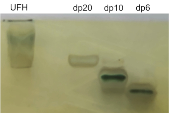
Figure 1: Silver stain development process. Alcian blue staining of unfractionated heparin (UFH) or size-defined oligosaccharides (dp = degree of polymerization) separated by electrophoresis is amplified by silver staining. From left to right: UFH, dp20, dp10, dp6. Please click here to view a larger version of this figure.
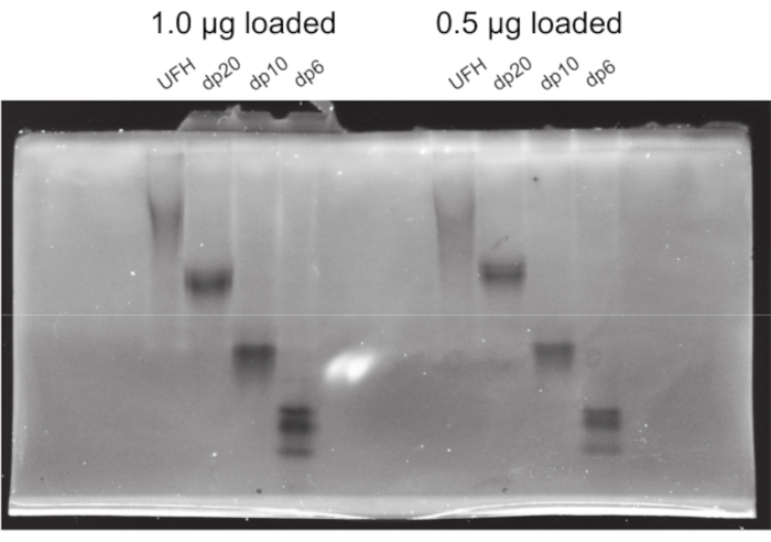
Figure 2: Sensitivity of polyacrylamide gel and silver stain for the detection for heparan sulfate. Heparan sulfate oligosaccharides of different lengths (unfractionated heparin aka UFH, dp20, dp10, dp6) were onto a 22% polyacrylamide gel and ran and silver stained. Each oligosaccharide was loaded twice at two different masses: 1.0 µg (leftmost bands) and 0.5 µg (rightmost bands). Please click here to view a larger version of this figure.
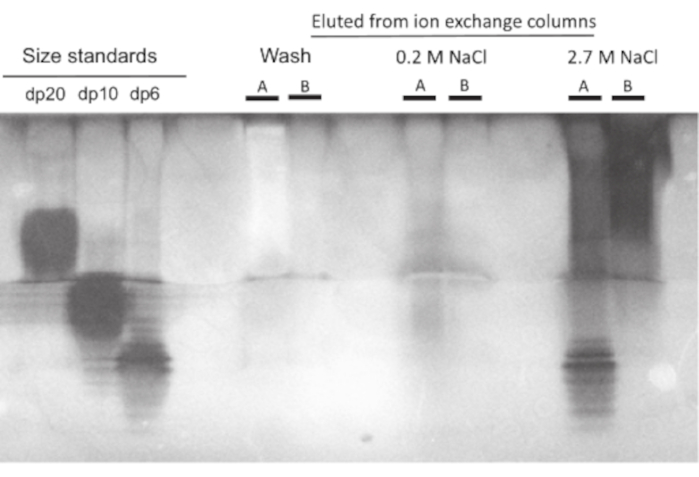
Figure 3: Efficiency of GAG purification from liquid biological samples. GAGs isolated from two bronchoalveolar lavage (BAL) samples, labeled here as "A" and "B", were run on a 22% polyacrylamide gel and silver stained. Sample A consisted of 1mL of BAL fluid harvested from a mouse 24 h after treatment with 3 mg/kg intratracheal lipopolysaccharide (LPS). An additional 10 µg of dp6 HS were added to this sample as a "spike in" control. Sample B consisted of 10 mL of pooled BAL fluid collected from 3 mice 24 h after administration of LPS. Each sample was run alongside fractions from the ion exchange column eluted with either diluent ("wash") or low concentrations of NaCl (0.2 M). 2 µg of commercially purchased dp20, dp10, and dp6 heparan sulfate oligosaccharides were used as size references (leftmost bands). Please click here to view a larger version of this figure.
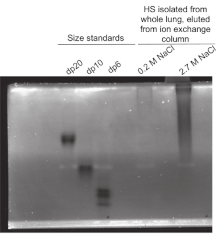
Figure 4: Measuring the size of heparan sulfates isolated and purified from a healthy mouse lung. Heparin sulfate content isolated and purified from 15 mg frozen sample of a lung isolated from a healthy mouse and run on a 22% polyacrylamide gel. Heparan sulfate was eluted from the ion exchange column with either low concentrations (0.2 M; low affinity fraction) or high concentrations (2.7 M, 16% solution; high affinity fraction) of NaCl. 2 µg of commercially purchased dp20, dp10, and dp6 heparan sulfate oligosaccharides were used as size references (leftmost bands). Please click here to view a larger version of this figure.
| NOTE: All solutions must be filtered (0.22μm) before use. | |||
| Solution | Recipe | Composition | Comments |
| Resolving gel/lower chamber running buffer (2 L or 4 L) | Boric acid, MW 61.83 | 2L: 12.36 g; 4L: 24.76 g | Desired concentration: 0.1 M |
| Tris base, MW 124.14 | 2L: 24.2 g; 4L: | Desired concentration: 0.1 M | |
| Disodium EDTA MW 336.21 or dihydrate, MW 372.36 | 2L: 6.7 g or 7.4 g, respectively; 4L 13.4 g or 14.8 g, respectively | Desired concentration: 0.01 M | |
| Deionized water | 2L or 4L | Adjust pH to 8.3 after fully dissolving reagents | |
| Upper chamber running buffer (1 L) | Glycine | 93 g | Desired concentration: 1 M |
| Tris base, MW 124.14 | 24.2 g | Desired concentration: 0.2 M | |
| Deionized water | 1 L | ||
| Resolving gel, 22% total acrylamide (500mL) | Acrylamide, MW 71.08 | 100.1 g | Desired concentration: 20.02% w/v |
| N,N'-methylene-bis-acrylamide (bis, MW 154.17) | 10 g | Desired concentration: 2% w/v | |
| Sucrose | 75 g | Desired concentration: 15% w/v | |
| Resolving gel buffer | 500 mL | Bring to a total volume of 500mL; will require less than 500mL of buffer total | |
| Resolving gel, 15% total acrylamide (400mL) | Acrylamide, MW 71.08 | 56.3 g | Desired concentration: 14.08% w/v |
| N,N'-methylene-bis-acrylamide (bis, MW 154.17) | 3.7 g | Desired concentration: 2% w/v | |
| Sucrose | 20.8 g | Desired concentration: 15% w/v | |
| Resolving gel buffer | 400 mL | Bring to a total volume of 400mL; will require less than 400mL of buffer total | |
| Stacking gel (100 mL) | Acrylamide, MW 71.08 | 4.75 g | Desired concentration: 4.75% w/v |
| N,N'-methylene-bis-acrylamide (bis, MW 154.17) | 0.25 g | Desired concentration: 0.25% w/v | |
| Resolving gel buffer | 100 mL | Add 80mL buffer and full dissolve reagents. Then adjust pH to 76.3 with hydrochloric acid, dropwise. Then bring to a total volume of 100 mL with resolving gel buffer. | |
| Sample loading buffer (500 mL) | Sucrose | 250g | Desired concentration: 50% w/v |
| Phenol red | 500mg | Desired concentration: 1mg/mL | |
| Bromophenol blue | 250mg | Desired concentration: 0.5mg/mL | |
| Deionized water | 500mL | Bring to a total volume of 500mL; will require less than 500mL of water total | |
| Heparin derived oligosacharide 'ladder' (2mL each) | dp6 | 0.1mg | NOTE: Recommend using heparin derived oligosaccharides 6 polymer subunits in length (aka degree of polymerization 6, or dp6) for the smallest band, dp20 for the largest band, and dp10 for middle band. However, other combinations can be used if desired. Desired concentration: 0.05 mg/mL |
| dp10 | 0.1mg | ||
| dp20 | 0.1mg | ||
| Deionized water | 3mL | Dissolve each oligosaccharide in separate tubes with 1mL each | |
| Sample loading buffer | 3mL | Add 1mL (1:1 mixture) to each oligosaccharide solution | |
Table 1: Solutions required for polyacrylamide gel electrophoresis of purified glycosaminoglycans. All solutions must be filtered (0.22 µm) before use.
| NOTE: All solutions must be filtered (0.22μm) before use. | |||
| Solution | Recipe | Composition | Comments |
| Alcian blue staining solution (500mL) | Alcian blue 8GX powder | 2.5g | Desired concentration: 0.5% w/v |
| 2% v/v glacial acetic acid | 500mL | ||
| Silver stain (200 mL) | Deionized water | 192.7 mL | Prepare stain solution fresh; use within one week. |
| 7.6M sodium hydroxide | 2 mL | To make, add 3.04 g sodium hydroxide pellets to 10 mL water | |
| Ammonium hydroxide | 3.3 mL | ||
| 4M silver nitrate solution | 2 mL | To make, add 3.397 g silver nitrate to 5 mL water. Add dropwise while stirring to avoid precipitation. | |
| Developing solution (501 mL) | Deionized water | 500 mL | Developing solution must be made fresh and used within 24 h. |
| 2.5% w/v citric acid | 1 mL | To make, add 100mg to 4 mL deionized water. Citric acid must be made fresh. | |
| Formaldehyde | 250μL | ||
| Stop solution (500 mL) | Deionized water | 300 mL | |
| Glacial acetic acid | 20 mL | ||
| Methanol | 90 mL | ||
Table 2: Solutions required for silver staining of glycosaminoglycans separated by polyacrlamide gel electrophoresis. All solutions must be filtered (0.22 µm) before use.
Discussion
GAGs play a central role in many diverse biological processes. One of the principal functions of sulfated GAGs (such as HS and CS) is to interact with and bind to ligands, which can alter downstream signaling functions. An important determinant of GAG binding affinity to cognate ligands is the length of the GAG polymer chain 8,9,14. For this reason, it is important for researchers to be able to define with reasonable precision the size of GAG chains isolated from biological samples of interest. To be practical, this technique should be capable of being performed using common laboratory equipment and reagents.
This protocol describes a method to isolate and purify GAGs from biological samples, to separate them by size via PAGE, and to visualize them using Alcian blue and silver staining technique. While there are several ways to separate glycosaminoglycans by size, our approach has several strengths specific to the application of this technique in life science laboratories. Firstly, with a limit of detection of 0.5 µg, this technique is highly sensitive in the detection of GAGs of interest. In our experience, even samples that contain relatively low concentrations of GAGs (i.e., BAL fluid samples) should yield more than enough GAG for detection using this method. While the yield from isolation and purification of BAL fluid will vary by technique and specific GAG of interest, it has been our experience that 10 mL of raw BAL fluid yields sufficient HS to be detectable using this technique. The yield from solid organs is substantially higher, depending on the tissue harvested, but it is our experience that an initial solid sample of 10 mg will yield ample HS for detection using this technique.
It is important to note that the final imaging of the stained PAGE gel will vary according to the imaging technologies available to the end-user. Digital photographs of the gel can be taken using a number of different systems, including numerous commercially available gel documentation systems or a regular commercial camera, depending the available equipment and the sensitivity of detection required (typically dictated by the amount of sample loaded onto the gel). It also should be noted that the light source required to digitally image this gel may vary depending on the density of the sample and the amount of development time required during the staining process. Gels that develop rapidly and produce silver-stained bands readily visible to the naked eye will require UV transillumination for the best images, as the majority of the light will be absorbed by unreacted Alcian blue. Gels that require longer development times (such as those with very small amounts of sample) will require either epi-illumination or transillumination with normal full spectrum light, as this will be best absorbed by the silver stain.
A further strength of this technique is that it is particularly adaptable to life sciences labs, due to its basis in simple PAGE technology, using equipment and reagents that are commonly available and cheaply acquired. While there are other approaches to quantifying the length of isolated GAG polymers (e.g., capillary electrophoresis), they typically require both knowledge and equipment that is not commonly available in most life sciences laboratories15,16. The simplicity of this approach and the relatively affordable and available nature of the reagents required makes this technique readily adaptable by life sciences researchers interested in studying GAG biology in the context of their given subfields. Furthermore, this technique serves as an essential complement to mass spectrometry-based techniques of detecting GAGs in biological tissues. While mass spectrometry based approaches are able to detect GAGs with high sensitivity and to discern subtle differences in structure from complex samples17, due to the nature of the technology it is not able to distinguish between GAG polymers by size. For this reason, a PAGE based approach is essential to divining the size of HS polymers in biological samples of interest.
It is important to note that there are several limitations to the techniques described in this paper. The first and most salient is that because of the charge-charge interaction that are central to the Alcian blue staining reaction, this approach is highly selective for highly sulfated GAGs and will only weakly stain more neutrally-charged moieties (e.g., hyaluronic acid18). Thus, this technique is likely to bias its results towards more acidic GAG moieties. For this reason, complementary approaches to measure GAG content in biological samples of interest (e.g., mass spectrometry-based techniques) should be used adjunctively to provide a more complete picture of the GAGs present in experimental samples. Furthermore, for end-users specifically interested in measuring the size of hyaluronan, others have described similar PAGE based techniques that leverage biotinylated hyaluronic acid binding protein (HABP) which may be adaptable to the basic technique described here19.
In summary, the technique presented in this article can be used to isolate, purify, and detect GAGs from biological samples with a great deal of sensitivity and specificity as well as to measure the native length of these polysaccharide chains. This information can be critical to testing hypotheses about GAG-ligand interactions due to the importance of GAG polymer length in determining cognate ligand binding affinity. This approach has several advantages, most notably its relative simplicity and adaptability to life sciences research laboratories, though it is limited by its relative bias towards negatively charged GAG moieties. Despite this drawback, this technique represents a robust tool that would encourage investigators to study the role of GAGs in organ homeostasis and disease.
Disclosures
The authors declare that they have no competing financial interests.
Acknowledgements
This work was funded by F31 HL143873-01 (WBL), R01 HL125371 (RJL and EPS)
Materials
| Name | Company | Catalog Number | Comments |
|---|---|---|---|
| Accuspin Micro17 benchtop microcentrifuge | thermoFisher Scientific | 13-100-675 | Any benchtop microcentrifuge/rotor combination capable of 14000 xG is appropriate |
| Acrylamide (solid) | thermoFisher Scientific | BP170-100 | Electrophoresis grade |
| Actinase E | Sigma Aldrich | P5147 | Protease mix from S. griseus |
| Alcian Blue 8GX (solid) | thermoFisher Scientific | AC400460100 | |
| Ammonium acetate (solid) | thermoFisher Scientific | A639-500 | Molecular biology grade |
| Ammonium hydroxide (liquid) | thermoFisher Scientific | A669S-500 | certified ACS |
| Ammonium persulfate (solid) | thermoFisher Scientific | BP179-25 | electrophoresis grade |
| Barnstead GenPure Pro Water Purification System | ThermoFisher Scientific | 10-451-217PKG | Any water deionizing/ purification system is an acceptable substitute |
| Boric acid (solid) | thermoFisher Scientific | A73-500 | Molecular biology grade |
| Bromphenol blue (solid) | thermoFisher Scientific | B392-5 | |
| Calcium acetate (solid) | ThermoFisher Scientific | 18-609-432 | Molecular biology grade |
| Calcium chloride (solid) | ThermoFisher Scientific | AC349610250 | Molecular biology grade |
| CHAPS detergent (3-((3-cholamidopropyl) dimethylammonio)-1-propanesulfonate) | ThermoFisher Scientific | 28299 | |
| Chondroitinase ABC | Sigma Aldrich | C3667 | |
| Criterion empty cassette for PAGE (1.0mm thick, 12+2 wells) | Bio-Rad | 3459901 | Any 1.0mm thick PAGE casting cassette system will suffice |
| Criterion PAGE Cell system (cell and power supply) | Bio-Rad | 1656019 | any comparable vertical gel PAGE system will work) |
| Dichloromethane (liquid) | thermoFisher Scientific | AC610931000 | certified ACS |
| EDTA disodium salt (solid) | thermoFisher Scientific | 02-002-786 | Molecular biology grade |
| Glacial acetic acid (liquid) | thermoFisher Scientific | A35-500 | Certified ACS |
| Glycine (solid) | thermoFisher Scientific | G48-500 | Electrophoresis grade |
| Heparanase I/III | Sigma Aldrich | H3917 | From Flavobacterium heparinum |
| Heparin derived decasaccharide (dp10) | galen scientific | HO10 | |
| Heparin derived hexasaccharde (dp6) | Galen scientific | HO06 | |
| Heparin derived oligosaccharide (dp20) | galen scientific | HO20 | |
| Hydrochloric acid (liquid) | thermoFisher Scientific | A466-250 | |
| Lyophilizer | Labconco | 7752020 | Any lyophilizer that can achieve -40C and 0.135 Torr will work; can also be replaced with rotational vacuum concentrator |
| Methanol (liquid) | thermoFisher Scientific | A412-500 | Certified ACS |
| Molecular Imager Gel Doc XR System | Bio-Rad | 170-8170 | Any comparable gel imaging system is an acceptable substitute |
| N,N'-methylene-bis-acrylamide (solid) | thermoFisher Scientific | BP171-25 | Electrophoresis grade |
| Phenol red (solid) | thermoFisher Scientific | P74-10 | Free acid |
| Q Mini H Ion Exchange Column | Vivapure | VS-IX01QH24 | Ion exchange column must have minimum loading volume of 0.4mL, working pH of 2-12, and selectivity for ionic groups with pKa of 11 |
| Silver nitrate (solid) | thermoFisher Scientific | S181-25 | certified ACS |
| Sodium Acetate (solid) | ThermoFisher Scientific | S210-500 | Molecular biology grade |
| Sodium chloride (solid) | thermoFisher Scientific | S271-500 | Molecular biology grade |
| Sodium hydroxide (solid) | thermoFisher Scientific | S392-212 | |
| Sucrose (solid) | thermoFisher Scientific | BP220-1 | Molecular biology grade |
| TEMED (N,N,N',N'-tetramethylenediamine) | thermoFisher Scientific | BP150-20 | Electrophoresis grade |
| Tris base (solid) | thermoFisher Scientific | BP152-500 | Molecular biology grade |
| Ultra Centrifugal filters, 0.5mL, 3000 Da molecular weight cutoff | Amicon | UFC500324 | Larger volume filter units may be used, depending on sample size. |
| Urea (solid) | ThermoFisher Scientific | 29700 | |
| Vacufuge Plus | Eppendorf | 22820001 | Any rotational vacuum concentrator will work; can be replaced with lyophilizer |
| Vacuum filter unit, single use, 0.22uM pore PES, 500mL volume | thermoFisher Scientific | 569-0020 | Alternative volumes and filter materials acceptable |
References
- LaRivière, W. B., Schmidt, E. P. The pulmonary endothelial glycocalyx in ARDS: A critical role for heparan sulfate. Current Topics in Membrane. 82, 33-52 (2018).
- Haeger, S. M., Yang, Y., Schmidt, E. P. Heparan sulfate in the developing, healthy, and injured lung. American Journal of Respiratory Cell and Molecular Biology. 55 (1), 5-11 (2016).
- Morita, H., Yoshimura, A., Kimata, K. The role of heparan sulfate in the glomerular basement membrane. Kidney International. 73 (3), 247-248 (2008).
- Schmidt, E. P., et al. The pulmonary endothelial glycocalyx regulates neutrophil adhesion and lung injury during experimental sepsis. Nature Medicine. 18 (8), 1217-1223 (2012).
- Haeger, S. M., et al. Epithelial heparan sulfate contributes to alveolar barrier function and is shed during lung injury. American Journal of Respiratry Cell and Molecular Biology. 59 (3), 363-374 (2018).
- Mankin, H. J., Lippiello, L. The glycosaminoglycans of normal and arthritic cartilage. Journal of Clinical Investigation. 50 (8), 1712-1719 (1971).
- Annaval, T., et al. Heparan sulfate proteoglycans biosynthesis and post synthesis mechanisms combine few enzymes and few core proteins to generate extensive structural and functional diversity. Molecules. 25 (18), (2020).
- Zhang, F., et al. Comparison of the interactions of different growth factors and glycosaminoglycans. Molecules. 24 (18), (2019).
- Pempe, E. H., Xu, Y., Gopalakrishnan, S., Liu, J., Harris, E. N. Probing structural selectivity of synthetic heparin binding to Stabilin protein receptors. Journal of Biological Chemistry. 287 (25), 20774-20783 (2012).
- Cowman, M. K., et al. Polyacrylamide-gel electrophoresis and Alcian Blue staining of sulphated glycosaminoglycan oligosaccharides. Biochemical Journal. 221 (3), 707-716 (1984).
- Møller, H. J., Poulsen, J. H. Improved method for silver staining of glycoproteins in thin sodium dodecyl sulfate polyacrylamide gels. Analytical Biochemistry. 226 (2), 371-374 (1995).
- Min, H., Cowman, M. K. Combined alcian blue and silver staining of glycosaminoglycans in polyacrylamide gels: Application to electrophoretic analysis of molecular weight distribution. Analytical Biochemistry. 155 (2), 275-285 (1986).
- Jay, G. D., Culp, D. J., Jahnke, M. R. Silver staining of extensively glycosylated proteins on sodium dodecyl sulfate-polyacrylamide gels: Enhancement by carbohydrate-binding dyes. Analytical Biochemistry. 185 (2), 324-330 (1990).
- Abraham, E., et al. Liposomal prostaglandin E1 (TLC C-53) in acute respiratory distress syndrome: a controlled, randomized, double-blind, multicenter clinical trial. TLC C-53 ARDS Study Group. Critical Care Medicine. 27 (8), 1478-1485 (1999).
- Pervin, A., al-Hakim, A., Linhardt, R. J. Separation of glycosaminoglycan-derived oligosaccharides by capillary electrophoresis using reverse polarity. Analytical Biochemistry. 221 (1), 182-188 (1994).
- Wang, Z., Zhang, F., Dordick, J. S., Linhardt, R. J. Molecular mass characterization of glycosaminoglycans with different degrees of sulfation in bioengineered heparin process by size exclusion chromatography. Current Analytical Chemistry. 8 (4), 506-511 (2012).
- Pepi, L. E., Sanderson, P., Stickney, M., Amster, I. J. Developments in mass spectrometry for glycosaminoglycan analysis: A review. Molecular and Cellular Proteomics. , 100025(2021).
- Whiteman, P. The quantitative measurement of Alcian Blue-glycosaminoglycan complexes. Biochemical Journal. 131 (2), 343-350 (1973).
- Yuan, H., et al. Molecular mass dependence of hyaluronan detection by sandwich ELISA-like assay and membrane blotting using biotinylated hyaluronan binding protein. Glycobiology. 23 (11), 1270-1280 (2013).
Reprints and Permissions
