Developmental Biology
A subscription to JoVE is required to view this content.
You will only be able to see the first 2 minutes.
The JoVE video player is compatible with HTML5 and Adobe Flash. Older browsers that do not support HTML5 and the H.264 video codec will still use a Flash-based video player. We recommend downloading the newest version of Flash here, but we support all versions 10 and above.
If that doesn't help, please let us know.
Visualization of Cell Cycle Variations and Determination of Nucleation in Postnatal Cardiomyocytes
Chapters
Summary February 24th, 2017
To distinguish cell division from cell cycle variations in cardiomyocytes, we present protocols using two transgenic mouse lines: Myh6-H2B-mCh transgenic mice, for the unequivocal identification of cardiomyocyte nuclei, and CAG-eGFP-anillin mice, for distinguishing cell division from cell cycle variations.
Transcript
The overall goal of this procedure is to visualize cell cycle activity in postnatal cardiomyocytes and to determine their nuclearity. This method can help answer key questions in the cardiac regeneration field, such as does a cardiomyocyte really divide or does it just increase its ploidy or become multi-nucleated? The main advantages of this technique are that the M phase of the cell cycle can be visualized in high spatiotemporal resolution and that cardiomyocyte nuclei can be identified by fluorescence.
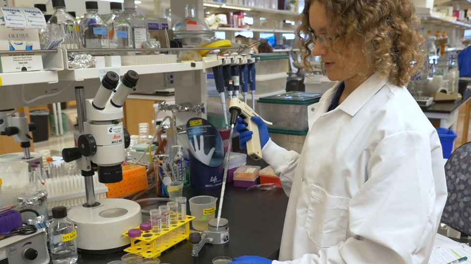
Demonstrating the procedure will be Patricia Freitag, a technician from our laboratory. If using non-homozygous breeding pairs, first identify the transgenic hearts via fluorescent microscopy to check for their EGFP analin and H2B-mCherry expression. EGFP analyin nuclear signals can be most easily detected at the edges of the atria by focusing through its tissue layers.
To isolate the cardiomyocytes, place the appropriate number of hearts in 1.5 milliliter reaction tubes containing one milliliter of enzyme mix from a neonatal heart dissociation kit and cut the hearts into small pieces with scissors. Then, transfer the solution into 15 milliliter reaction tubes. Place the tube in an almost horizontal position at 37 degrees celsius for 15 minutes, mixing the tissue five to 10 times with a five milliliter pipette at the end of the incubation.
At the end of the third digestion, stop the reaction with 7.5 milliliters of medium two and filter the cell suspension through a 70 micron cell strainer. Rinse the cell strainer with three milliliters of medium two, pooling the wash with the collected cells and collect the cells by centrifugation. Re-suspend the pellet in 500 microliters of medium one for counting.
Then, seed one times ten to the fourth cells per well in 120 microliters of medium one in a 96-well flat-bottom plate and centrifuge the cells before their overnight culture in a cell culture incubator. The next day, most of the cardiomyocytes should be beating. Before beginning the transfection, wipe the bench and all of the transfection materials with RNAse decontamination solution to remove any RNAse.
When all of the materials are ready, add 20 microliters of freshly prepared siRNA mix to each well and return the cells to the incubator for 48 hours. After two days, replace the supernatant in each well with 120 microliters of medium one and return the cells to the incubator for at least another 24 hours. At the end of the incubation, wash the cells one time with PBS, followed by fixation in 4%formaldehyde solution for 20 minutes.
To analyze the cells by confocal video microscopy, transfer the plate to the confocal microscope stage and start the imaging software. Open the A1plus settings and check the channel one, two, three and four boxes. In the pull-down menu, set channel one to DAPI, channel two to eGFP, channel three to RFP, and channel four to Cy5.
Click the channel voltage and use the slide bars to set the voltage to 80 for each channel. Use the slide bars to set the offset to zero and click on the home button to set the pinhole to the home position. Set the scan size to 1024 by 1024 pixels in the pull-down menu.
Click optimize to open the XYZ size setup window and check the perfect voxel box under the suggested step Z.Select the 20x objective and adjust the laser intensities until the picture is neither under-nor overexposed. When all of the parameters have been set, open the acquire menu. Select scan large image then choose current position is at top left corner under area.
Then set the number of fields in X and Y to three by three and click scan. To define the number of cardiomyocyte nuclei, click measure, manual measurement, and counts. Select the nuclei with H2B-mCherry signals, thereby marking and counting them.
Then, select the alpha-actinin positive cells to determine the number of cardiomyocytes. To count the G1, S, G2 phase cardiomyocytes with nuclear EGFP analin signals, select measure, manual measurement, and counts. Mark the EGFP analin and H2B-mCherry expressing nuclei with clicking and manually count the cardiomyocytes with the mitosis-specific EGFP analin signals such as contractile rings and mid-bodies.
Then, click measure, manual measurement, and counts and click the cardiomyocytes with mitosis-specific EGFP analin signals, cytoplasmatic signals, contractile rings, and mid-bodies. Compared to the negative controls, the miRNA and siRNA transfected cardiomyocytes demonstrate an induction of cell cycle activity. Cardiomyocytes that perform endoreduplication exhibit an exclusively nuclear EGFP analin expression, whereas cardiomyocytes undergoing cell division display EGFP analin expression in M phase typical localizations.
After enzymatic digestion of the heart tissue at the Langendorff apparatus, the atria and ventricles can be mechanically separated and analyzed independently, allowing the visualization of H2B-mCherry transgenic ventricular cardiomyocytes with a high number of H2B-mCherry positive bi-nucleated cardiomyocytes. By contrast, the majority of atrial cardiomyocytes are mono-nucleated. Enzymatic digestion does not result in a 100%single-cell population revealing a pattern of cross-striation by alpha-actinin staining, further facilitating the discrimination between bi-nucleated cardiomyocytes as a continuous pattern of cross-striation and cell doublets.
To analyze the bi-nucleation index of cardiomyocytes in thick slices, it is necessary to manually scroll through the stack as the nuclei do not necessarily lay within one Z-plane, with wheat germ agglutinin staining permitting the detection of the cell borders. 3D reconstructions of fixed slices of adult transgenic mouse hearts allow the detection and automated counting of hooks, stained, and H2B-mCherry positive nuclei to determine the proportion of cardiomyocyte nuclei under physiological conditions within the tissue. Once mastered, the heart dissociation can be completed in two to three hours if it is performed properly.
While attempting this procedure, it is important to remember to work continuously to maintain the viability of the cells throughout the experiment. Following this procedure, it is possible to screen microRNAs or other small substances for their proliferation-inducing effects in postnatal cardiomyocytes.
Related Videos
You might already have access to this content!
Please enter your Institution or Company email below to check.
has access to
Please create a free JoVE account to get access
Login to access JoVE
Please login to your JoVE account to get access
We use/store this info to ensure you have proper access and that your account is secure. We may use this info to send you notifications about your account, your institutional access, and/or other related products. To learn more about our GDPR policies click here.
If you want more info regarding data storage, please contact gdpr@jove.com.
Please enter your email address so we may send you a link to reset your password.
We use/store this info to ensure you have proper access and that your account is secure. We may use this info to send you notifications about your account, your institutional access, and/or other related products. To learn more about our GDPR policies click here.
If you want more info regarding data storage, please contact gdpr@jove.com.
Your JoVE Unlimited Free Trial
Fill the form to request your free trial.
We use/store this info to ensure you have proper access and that your account is secure. We may use this info to send you notifications about your account, your institutional access, and/or other related products. To learn more about our GDPR policies click here.
If you want more info regarding data storage, please contact gdpr@jove.com.
Thank You!
A JoVE representative will be in touch with you shortly.
Thank You!
You have already requested a trial and a JoVE representative will be in touch with you shortly. If you need immediate assistance, please email us at subscriptions@jove.com.
Thank You!
Please enjoy a free 2-hour trial. In order to begin, please login.
Thank You!
You have unlocked a 2-hour free trial now. All JoVE videos and articles can be accessed for free.
To get started, a verification email has been sent to email@institution.com. Please follow the link in the email to activate your free trial account. If you do not see the message in your inbox, please check your "Spam" folder.