8,289 Views
•
08:24 min
•
May 22, 2017
DOI:
The overall goal of this article is to introduce preparation and characterization methods for nerve growth factor-loaded, high-density lipoprotein-mimicking nanoparticles. Simple homogenization was used to prepare normal, high-density lipoprotein-mimicking nanoparticles. The procedure is simple and scalable.
Moreover, the nanoparticle greatly improved entrapment efficiency, also NGF, and APOA1 in the nanoparticles. Various challenges to characterize a nanoparticle were overcome, including separate to the unloaded NGF from the NGF nanoparticles, conduct reliable in vitro radius studies, and the bioavailability of NGF nanoparticles. To begin, mix 10 microliters of NGF with 10 microliters of protamine USP in a 1.5 milliliter microcentrifuge tube and let it stand for 10 minutes at room temperature to form the complex.
Next, add 59 microliters of phosphatidylcholine and 11 microliters of sphingomyelin to a glass vial. Also add four microliters of phosphatidylserine, 15 microliters of cholesteryl oleate, and 45 microliters of tocopheryl polyethylene glycol succinate. Mix and evaporate the ethanol under a gentle nitrogen stream for about five minutes.
All excipients should form an oily thin film at the bottom of the glass vial. Add one milliliter of ultrapure water to the vial and homogenize at 9, 500 rpm for five minutes at room temperature to form the prototype nanoparticles, or NPs. Then, add the prepared complex to the prototype NPs.
Incubate the suspension at 37 degrees Celsius for 30 minutes with stirring, by using a small stirring bar in the glass vial. Cool the NPs down by stirring at room temperature for another 30 minutes. After cooling, add 106 microliters or apolipoprotein A-1 and stir at room temperature overnight to form the final NGF HDL-mimicking NPs.
Measure the particle size and zeta potential using a particle analyzer as per the manufacturer’s instructions. A cross-linked gel filtration chromatographic column is used to separate the unloaded NGF from the NGF HDL-mimicking nanoparticles and determines entrapment efficiency of the NGF. For the column preparation, transfer 15 milliliters of sepharose 4B CL suspension to a 50 milliliter beaker.
Stir the bead suspension and pour some into a column. Gently tap the column to get rid of bubbles. Allow the solvent to drain and the beads to settle for a few minutes.
Continue to add the remaining suspension. Rinse the inside wall of the column to clean the beads. Drain the solvent until the solvent level is slightly above the top of the stationary phase.
Then, wash and condition the column with 20 milliliters of 1X phosphate-buffered saline. To measure the entrapment efficiency of the NGF, load 200 microliters of NGF HDL-mimicking NPs onto the column and elute with 1X PBS. As before, collect a total of 12 one-milliliter fractions.
Pretreat eight dialysis tubes by following the manufacturer’s instructions. Add 30 milliliters of release medium to a 50 milliliter centrifuge tube. Then, warm it up to 37 degrees Celsius in a shaker with a 135 rpm shaking speed.
Close the dialysis tube and place it into the release medium. Quickly withdraw 100 microliters of the release medium from outside the dialysis tube as the sample for time zero. Immediately put the sample into minus 20 degrees Celsius for later analysis.
Add 100 microliters of fresh release medium into the centrifuge tube to replace the withdrawn sample. Then, start the timer. At 1, 2, 4, 6, 8, 24, 48, and 72 hours, withdraw 100 microliters of the release medium and replace it with 100 microliters of fresh medium.
Immediately put the withdrawn samples into minus 20 degrees Celsius for later analysis. After a three-minute homogenization, consistent particle sizes were obtained for the prototype nanoparticles. The final NGF nanoparticles had particle sizes of 170 nanometers with a narrow and monodispersed size distribution.
Unloaded NGF and nanoparticles were completely separated on the gel filtration column. Nanoparticles were eluded in fractions two through four. ELISA measurement indicated that free NGF was eluded in fraction 7 to fraction 10.
The calculated entrapment efficiency of NGF was approximately 66%In vitro release of NGF nanoparticles is shown here. By adding PBS and BSA to the release medium, free NGF passed through the dialysis membrane with a recovery of over 85%Conversely, NGF nanoparticles showed a slow release profile with about 10%of NGF being released from the nanoparticles over 72 hours. NGF nanoparticles stimulated neurite outgrowth in PC12 cells to a similar extent as for free NGF.
Neurite outgrowth was clearly observed under the microscope for both groups when dosed at 50 nanograms per milliliter. NGF nanoparticles considerably increased the plasma concentration and decreased the uptake of NGF in the mice liver, kidney, and spleen. HDL-mimicking nanoparticles assisted NGF, allowing for a long half-life in in vivo stability.
Once mastered, monodispersed nanoparticles can be prepared in three minutes using a homogenization method if it is performed properly. While attempting this procedure, it is important to remember to use a gentle nitrogen stream to evaporate to the solvent and make sure no lipid residue sticking on the wall of the container after homogenization. After watching this video, you should have a good understanding of how to make HDL-mimicking nanoparticles using a homogenization method and how to characterize NGF loaded nanoparticles.
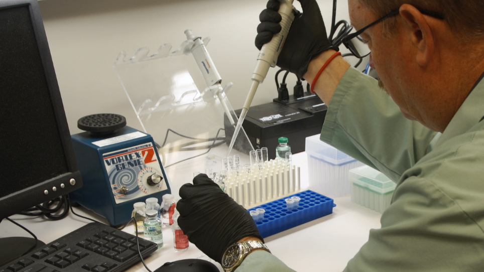
Simple homogenization was used to prepare novel, high-density, lipoprotein-mimicking nanoparticles to encapsulate nerve growth factor. Challenges, detailed protocols for nanoparticle preparation, in vitro characterization, and in vivo studies are described in this article.
08:35
Therapeutic Gene Delivery and Transfection in Human Pancreatic Cancer Cells using Epidermal Growth Factor Receptor-targeted Gelatin Nanoparticles
Related Videos
27999 Views
09:29
Electrospinning Growth Factor Releasing Microspheres into Fibrous Scaffolds
Related Videos
12208 Views
11:11
Construction and Characterization of a Novel Vocal Fold Bioreactor
Related Videos
9106 Views
09:16
Studying the Stoichiometry of Epidermal Growth Factor Receptor in Intact Cells using Correlative Microscopy
Related Videos
8327 Views
12:27
Polyelectrolyte Complex for Heparin Binding Domain Osteogenic Growth Factor Delivery
Related Videos
7510 Views
09:56
Preparation and Characterization of Lipophilic Doxorubicin Pro-drug Micelles
Related Videos
14818 Views
10:12
Flash NanoPrecipitation for the Encapsulation of Hydrophobic and Hydrophilic Compounds in Polymeric Nanoparticles
Related Videos
21797 Views
06:15
Detection of Endotoxin in Nano-formulations Using Limulus Amoebocyte Lysate (LAL) Assays
Related Videos
14405 Views
10:12
Designing Porous Silicon Films as Carriers of Nerve Growth Factor
Related Videos
9679 Views
09:49
Growth and Characterization of Irradiated Organoids from Mammary Glands
Related Videos
8021 Views
Read Article
Cite this Article
Zhu, J., Dong, X. Preparation and Characterization of Novel HDL-mimicking Nanoparticles for Nerve Growth Factor Encapsulation. J. Vis. Exp. (123), e55584, doi:10.3791/55584 (2017).
Copy