-1::1
Simple Hit Counter
Skip to content

Products

Solutions

×
×
Sign In

EN

EN - EnglishCN - 简体中文DE - DeutschES - EspañolKR - 한국어IT - ItalianoFR - FrançaisPT - Português do BrasilPL - PolskiHE - עִבְרִיתRU - РусскийJA - 日本語TR - TürkçeAR - العربية
Sign In Start Free Trial

RESEARCH

JoVE Journal

Peer reviewed scientific video journal

Behavior
Biochemistry
Bioengineering
Biology
Cancer Research
Chemistry
Developmental Biology
View All
JoVE Encyclopedia of Experiments

Video encyclopedia of advanced research methods

Biological Techniques
Biology
Cancer Research
Immunology
Neuroscience
Microbiology
JoVE Visualize

Visualizing science through experiment videos

EDUCATION

JoVE Core

Video textbooks for undergraduate courses

Analytical Chemistry
Anatomy and Physiology
Biology
Cell Biology
Chemistry
Civil Engineering
Electrical Engineering
View All
JoVE Science Education

Visual demonstrations of key scientific experiments

Advanced Biology
Basic Biology
Chemistry
View All
JoVE Lab Manual

Videos of experiments for undergraduate lab courses

Biology
Chemistry

BUSINESS

JoVE Business

Video textbooks for business education

Accounting
Finance
Macroeconomics
Marketing
Microeconomics

OTHERS

JoVE Quiz

Interactive video based quizzes for formative assessments

Authors

Teaching Faculty

Librarians

K12 Schools

Products

RESEARCH

JoVE Journal

Peer reviewed scientific video journal

JoVE Encyclopedia of Experiments

Video encyclopedia of advanced research methods

JoVE Visualize

Visualizing science through experiment videos

EDUCATION

JoVE Core

Video textbooks for undergraduates

JoVE Science Education

Visual demonstrations of key scientific experiments

JoVE Lab Manual

Videos of experiments for undergraduate lab courses

BUSINESS

JoVE Business

Video textbooks for business education

OTHERS

JoVE Quiz

Interactive video based quizzes for formative assessments

Solutions

Authors
Teaching Faculty
Librarians
K12 Schools

Language

English

EN

English

CN

简体中文

DE

Deutsch

ES

Español

KR

한국어

IT

Italiano

FR

Français

PT

Português do Brasil

PL

Polski

HE

עִבְרִית

RU

Русский

JA

日本語

TR

Türkçe

AR

العربية

    Menu

    JoVE Journal

    Behavior

    Biochemistry

    Bioengineering

    Biology

    Cancer Research

    Chemistry

    Developmental Biology

    Engineering

    Environment

    Genetics

    Immunology and Infection

    Medicine

    Neuroscience

    Menu

    JoVE Encyclopedia of Experiments

    Biological Techniques

    Biology

    Cancer Research

    Immunology

    Neuroscience

    Microbiology

    Menu

    JoVE Core

    Analytical Chemistry

    Anatomy and Physiology

    Biology

    Cell Biology

    Chemistry

    Civil Engineering

    Electrical Engineering

    Introduction to Psychology

    Mechanical Engineering

    Medical-Surgical Nursing

    View All

    Menu

    JoVE Science Education

    Advanced Biology

    Basic Biology

    Chemistry

    Clinical Skills

    Engineering

    Environmental Sciences

    Physics

    Psychology

    View All

    Menu

    JoVE Lab Manual

    Biology

    Chemistry

    Menu

    JoVE Business

    Accounting

    Finance

    Macroeconomics

    Marketing

    Microeconomics

Start Free Trial
Loading...
Home
JoVE Journal
Bioengineering
Autologous Endothelial Progenitor Cell-Seeding Technology and Biocompatibility Testing For Cardio...
JoVE Journal
Bioengineering
This content is Free Access.
JoVE Journal Bioengineering
Autologous Endothelial Progenitor Cell-Seeding Technology and Biocompatibility Testing For Cardiovascular Devices in Large Animal Model

Autologous Endothelial Progenitor Cell-Seeding Technology and Biocompatibility Testing For Cardiovascular Devices in Large Animal Model

Full Text
20,763 Views
11:49 min
September 9, 2011

DOI: 10.3791/3197-v

Alexandra E. Jantzen1, Whitney O. Lane2, Shawn M. Gage3, Justin M. Haseltine1, Lauren J. Galinat1, Ryan M. Jamiolkowski4, Fu-Hsiung Lin3, George A. Truskey1, Hardean E. Achneck3

1Department of Biomedical Engineering,Duke University , 2School of Medicine,Duke University , 3Department of Surgery,Duke University Medical Center, 4School of Medicine,University of Pennsylvania

Summary

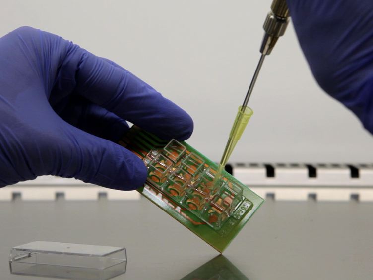
A method for seeding titanium blood-contacting biomaterials with autologous cells and testing biocompatibility is described. This method uses endothelial progenitor cells and titanium tubes, seeded within minutes of surgical implantation into porcine venae cavae. This technique is adaptable to many other implantable biomedical devices.

Transcript

The overall goal of this experiment is to seed vascular implants with autologous blood derived endothelial Progenitor cells then observe the effect of this treatment on thrombosis in a large animal model. This is achieved by obtaining a peripheral blood sample from swine to isolate late outgrowth porcine endothelial progenitor cells. Next, an implantable device.

The titanium tube in our proof of principle study is seated with the autologous PCs. The device is then implanted into the pig's inferior vena cava. Results show there's protection against thrombosis based on patency of the EPC seated titanium tube as opposed to the control and occluded bare metal tube.

Duke surgery is committed to fostering groundbreaking, innovative research. The following presentation is one example of how we work across departments and disciplines to advance medicine. Dr.EK has been leading a research effort to develop a cell seating technology that will prevent thrombosis by lining the blood contacting surface of vascular devices with patient's own progenitor cells.

Since these cells have been derived from the same patients that will receive the implant, we avoid rejection by the immune system. Our success and the methods described herein are the results of a large team effort. The surgical implantation will be demonstrated by Sean Gage from the section of vascular surgery, Whitney Lane, a Duke medical student, and myself.

I would now like to introduce Alexander Janssen, a PhD student from the Department of Biomedical Engineering, who will take us through the cell seeding process In detail, I will demonstrate how to seed titanium tubes with blood derived endothelial progenitor cells just prior to implantation into a pig model. This method serves as proof of concept and the same basic principles can be applied to many other implantable devices such as nitinol stents and mechanical circulatory assist devices. EPCs are isolated by plating the mononuclear cell fraction of porcine blood and usually appear after about seven days.

They can be recognized by their typical cobblestone morphology. Expand EPCs to a total number of 9 million cells in culture. Rinse the prepared EPCs twice in serum free medium, taking care to limit their light exposure during and after labeling.

Then cover the cells with a fluorescent label, PKH 26 and record how much solution is applied. Incubate the cells at room temperature for four minutes. Stop the labeling reaction by adding an equal volume of porcine serum to the dye solution one minute later.

Dilute the combined solutions one-to-one with full growth medium. Then aspirate the liquid and rinse the cells three times with full growth medium followed by two washes and DPBS without calcium or magnesium chloride. Next, cover the cells in trypsin taking note of the volume used and incubate them at 37 degrees Celsius.

After three minutes of incubation, use microscopy to check that the cells have detached, and if so, add twice as much trypsin neutralization solution as was added. Trypsin, combine the cell solutions into a single tube and measure the cell density with a hemo cytometer. Lastly, pellet the cell suspension and resuspend the cells in at least 4.5 milliliters of serum free medium.

After preparing the pig for surgery, incise the midline with a number 15 scalpel blade from the second set of memory glands. Cran lead to the second to last set coddly using electrocautery. Carry the dissection down to the abdominal fascia.

Lift the peritoneum with mosquito forceps and carefully enter it with mets andal scissors, externalize the urinary bladder and place a three oh Vicryl purse string suture into the bladder wall. Make a stab incision at its center and insert a 16 French Foley catheter into the incision. Then administer intravenous fluids to titrate the urine output to at least one milliliter per kilogram per hour during the surgery.

Next, externalize the small enlarged bowel and place two bour surgical retractors to expose the posterior aspect of the peritoneal cavity and identify the IVC using sharp and blunt dissection. Carefully free the IVC from surrounding tissue. Then skeletonize the vessel from the right renal artery proximal to the bifurcation of the IVC distal exercise.

Extreme care during the IVC dissection as even a very small defect in the IVC may lead to rapid hemorrhage and exsanguination of the animal ligate. All of the side branches of the IVC segment take special care dissecting and ligating the two large posterior lumbar veins immediately proximal to the bifurcation of the IVC. There's often a large lumbar vein on the posterior side.

Dissect this vein free and control it with vessel loops. Now in parallel to the surgery, begin seeding the implant with EPCs prior to beginning this procedure. The titanium tube tissue seating scaffolds must be fully assembled and ready for use in a sterile field.

Open the gas sterilized titanium tube assembly in a five cc syringe. Then remove the plunger from the five cc syringe, but do not dispose of it A fixed allure cap to the syringe. Use a pipette to fill the syringe with the EPC suspension.

Then replace the plunger. Remove the cap and insert the lure end of the syringe into the open silicone tubing of the assembly until it is just snug. But no further remove any bubbles by slowly advancing the cell solution until it reaches the top of the headpiece.

Then close the assembly with a lure cap and tape it shut in a sterile glove. Insert this entire assembly into a machined syringe holder and place it in a 37 degree Celsius incubator. Use a gauge to ensure that the seating chambers level and allow the assembly to incubate with gentle rotation.

Continue the surgery just after ligating the IVC by administering 100 units of heparin per kilogram pig. Then immediately clamp the IVC at a 45 degree angle distally on the IVC and lumbar vein, and then proximally. Next, use a number 11 scalpel blade and pot scissors to make a four centimeter longitudinal vein otomy between the proximal and distal clamps.

Evacuate any blood from the IVC and flush its inner lumen with sterile DPBS. Now insert the EPC seated implant or a bare metal control into the IVC to prevent the cells from drying and to evacuate air. Fill the IVC with DPBS with calcium chloride and magnesium chloride.

Then close the vein otomy with a four oh proline running suture and remove one proximal clamp to de air the IVC through a small amount of back bleeding. After the bleed, remove the distal clamp to keep the implant in place. Apply a stay suture into the PVC tubing going through the vein wall.

Close the fascia with OPDS on a CT needle. Close the subcutaneous space with two o Vicryl running. Then staple the skin closed.

Administer up to 20 milliliters of 0.25%Marcaine subcutaneously along the incision site and cover the wound with gauze and tegaderm for pain. Give flu Nixon and oxymorphone subcutaneously as needed. Discontinue the anesthesia and when the pig awakens, return it to its cage over the next seven days.

Administer atri every 24 hours as an antibiotic prophylactic and be sure that the fentanyl patch applied prior to surgery stays adhered for a total of three days. After three days, the seated titanium tube implants were removed and the inside surfaces were examined. Under 550 nanometer excitation wavelength, a confluent layer of fluorescently labeled cells was found in another tube.

Section cells were additionally stained with a platelet endothelial cell adhesion marker to visualize cell borders. A wide variety of stains can be used in this procedure. By contrast, a bare titanium tube implant completely occludes in the prothrombotic low share environment of the inferior vena cava.

As illustrated in images A and C, the cell seated titanium tube is shown in image B and remained free of clot Following our say isolation procedure with peak blood. EPC appear after a average seven days in culture While performing the surgical procedure, it is very important to both carefully and gently skeletonize the IVC cause even the smallest tear in the IVC or its side branches can lead to exsanguination of the pig. Following this procedure, other blood contacting materials can be rapidly endothelial lies to prevent thrombosis.

Endothelial vascular stents, for example, with our EPCs mimics the native lining of the blood vessel wall and may prevent stent re-stenosis and thrombosis After its development. Our quick see technology will allow for lining intravascular devices with patients own blood derived endothelial progenitor cells within minutes in the operating room or catheterization laboratory. Therefore, prolonged exvivo culture.

Time on the device itself is avoided and the technology becomes practical for translation into clinical practice.

Explore More Videos

Autologous Endothelial Progenitor Cell-seeding TechnologyBiocompatibility TestingCardiovascular DevicesLarge Animal ModelImplantable DevicesArtificial MaterialsThromboemboli FormationTi TubesBlood-derived Human Or Porcine Endothelial Progenitor Cells (EPCs)Inferior Vena Cava (IVC)BiocompatibilityProthrombotic EnvironmentBare Metal SurfacesEndothelialize DevicesAntithrombotic FunctionPeripheral BloodMononuclear Cell FractionCultureLaparotomy

Related Videos

Embryonic Stem Cell-Derived Endothelial Cells for Treatment of Hindlimb Ischemia

09:11

Embryonic Stem Cell-Derived Endothelial Cells for Treatment of Hindlimb Ischemia

Related Videos

12K Views

Isolation and Large Scale Expansion of Adult Human Endothelial Colony Forming Progenitor Cells

12:17

Isolation and Large Scale Expansion of Adult Human Endothelial Colony Forming Progenitor Cells

Related Videos

11K Views

Bioengineering Human Microvascular Networks in Immunodeficient Mice

06:55

Bioengineering Human Microvascular Networks in Immunodeficient Mice

Related Videos

12.6K Views

Implantation of Inferior Vena Cava Interposition Graft in Mouse Model

12:39

Implantation of Inferior Vena Cava Interposition Graft in Mouse Model

Related Videos

12.2K Views

Surgical Technique for the Implantation of Tissue Engineered Vascular Grafts and Subsequent In Vivo Monitoring

11:17

Surgical Technique for the Implantation of Tissue Engineered Vascular Grafts and Subsequent In Vivo Monitoring

Related Videos

12K Views

Generation and Grafting of Tissue-engineered Vessels in a Mouse Model

13:04

Generation and Grafting of Tissue-engineered Vessels in a Mouse Model

Related Videos

12.3K Views

The Arteriovenous (AV) Loop in a Small Animal Model to Study Angiogenesis and Vascularized Tissue Engineering

08:53

The Arteriovenous (AV) Loop in a Small Animal Model to Study Angiogenesis and Vascularized Tissue Engineering

Related Videos

12.8K Views

A Pre-Clinical Porcine Model of Orthotopic Heart Transplantation

09:12

A Pre-Clinical Porcine Model of Orthotopic Heart Transplantation

Related Videos

9.8K Views

Transplantation of a 3D Bioprinted Patch in a Murine Model of Myocardial Infarction

07:47

Transplantation of a 3D Bioprinted Patch in a Murine Model of Myocardial Infarction

Related Videos

6.9K Views

Implantation of Tissue-Engineered Vascular Graft in Mouse Carotid Artery via Cuff Technique

07:13

Implantation of Tissue-Engineered Vascular Graft in Mouse Carotid Artery via Cuff Technique

Related Videos

568 Views

JoVE logo
Contact Us Recommend to Library
Research
  • JoVE Journal
  • JoVE Encyclopedia of Experiments
  • JoVE Visualize
Business
  • JoVE Business
Education
  • JoVE Core
  • JoVE Science Education
  • JoVE Lab Manual
  • JoVE Quizzes
Solutions
  • Authors
  • Teaching Faculty
  • Librarians
  • K12 Schools
About JoVE
  • Overview
  • Leadership
Others
  • JoVE Newsletters
  • JoVE Help Center
  • Blogs
  • Site Maps
Contact Us Recommend to Library
JoVE logo

Copyright © 2025 MyJoVE Corporation. All rights reserved

Privacy Terms of Use Policies
WeChat QR code