-1::1
Simple Hit Counter
Skip to content

Products

Solutions

×
×
Sign In

EN

EN - EnglishCN - 简体中文DE - DeutschES - EspañolKR - 한국어IT - ItalianoFR - FrançaisPT - Português do BrasilPL - PolskiHE - עִבְרִיתRU - РусскийJA - 日本語TR - TürkçeAR - العربية
Sign In Start Free Trial

RESEARCH

JoVE Journal

Peer reviewed scientific video journal

Behavior
Biochemistry
Bioengineering
Biology
Cancer Research
Chemistry
Developmental Biology
View All
JoVE Encyclopedia of Experiments

Video encyclopedia of advanced research methods

Biological Techniques
Biology
Cancer Research
Immunology
Neuroscience
Microbiology
JoVE Visualize

Visualizing science through experiment videos

EDUCATION

JoVE Core

Video textbooks for undergraduate courses

Analytical Chemistry
Anatomy and Physiology
Biology
Cell Biology
Chemistry
Civil Engineering
Electrical Engineering
View All
JoVE Science Education

Visual demonstrations of key scientific experiments

Advanced Biology
Basic Biology
Chemistry
View All
JoVE Lab Manual

Videos of experiments for undergraduate lab courses

Biology
Chemistry

BUSINESS

JoVE Business

Video textbooks for business education

Accounting
Finance
Macroeconomics
Marketing
Microeconomics

OTHERS

JoVE Quiz

Interactive video based quizzes for formative assessments

Authors

Teaching Faculty

Librarians

K12 Schools

Products

RESEARCH

JoVE Journal

Peer reviewed scientific video journal

JoVE Encyclopedia of Experiments

Video encyclopedia of advanced research methods

JoVE Visualize

Visualizing science through experiment videos

EDUCATION

JoVE Core

Video textbooks for undergraduates

JoVE Science Education

Visual demonstrations of key scientific experiments

JoVE Lab Manual

Videos of experiments for undergraduate lab courses

BUSINESS

JoVE Business

Video textbooks for business education

OTHERS

JoVE Quiz

Interactive video based quizzes for formative assessments

Solutions

Authors
Teaching Faculty
Librarians
K12 Schools

Language

English

EN

English

CN

简体中文

DE

Deutsch

ES

Español

KR

한국어

IT

Italiano

FR

Français

PT

Português do Brasil

PL

Polski

HE

עִבְרִית

RU

Русский

JA

日本語

TR

Türkçe

AR

العربية

    Menu

    JoVE Journal

    Behavior

    Biochemistry

    Bioengineering

    Biology

    Cancer Research

    Chemistry

    Developmental Biology

    Engineering

    Environment

    Genetics

    Immunology and Infection

    Medicine

    Neuroscience

    Menu

    JoVE Encyclopedia of Experiments

    Biological Techniques

    Biology

    Cancer Research

    Immunology

    Neuroscience

    Microbiology

    Menu

    JoVE Core

    Analytical Chemistry

    Anatomy and Physiology

    Biology

    Cell Biology

    Chemistry

    Civil Engineering

    Electrical Engineering

    Introduction to Psychology

    Mechanical Engineering

    Medical-Surgical Nursing

    View All

    Menu

    JoVE Science Education

    Advanced Biology

    Basic Biology

    Chemistry

    Clinical Skills

    Engineering

    Environmental Sciences

    Physics

    Psychology

    View All

    Menu

    JoVE Lab Manual

    Biology

    Chemistry

    Menu

    JoVE Business

    Accounting

    Finance

    Macroeconomics

    Marketing

    Microeconomics

Start Free Trial
Loading...
Home
JoVE Journal
Biochemistry
Mouse Adipose Tissue Collection and Processing for RNA Analysis
JoVE Journal
Biochemistry
A subscription to JoVE is required to view this content.  Sign in or start your free trial.
JoVE Journal Biochemistry
Mouse Adipose Tissue Collection and Processing for RNA Analysis

Mouse Adipose Tissue Collection and Processing for RNA Analysis

Full Text
51,247 Views
11:13 min
January 31, 2018

DOI: 10.3791/57026-v

Paul Tan1,2, Émilie Pepin1,4, Julie L. Lavoie1,3,4

1Centre de Recherche du Centre Hospitalier,Université de Montréal, 2Department of Biochemistry and Molecular Medicine,Université de Montréal, 3Department of Kinesiology,Université de Montréal, 4Montreal Diabetes Research Center

Summary

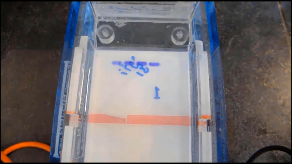
The purpose of this paper is to present a step-by-step procedure to collect different white adipose tissues from mice, process the fat samples and extract RNA.

Transcript

The overall goal of this procedure is to ensure adequate sample collection of white adipose tissue depots from different mice, and to isolate a high amount of good-quality RNA from the tissue. This method can answer key questions in the fields of metabolism, biochemistry and molecular biology that, for example, pertain to gene expression in adipose tissue from treated or genetically-modified mice. The main advantage of this technique is that it allows for the isolation of high amounts of good-quality RNA from adipose tissue which typically has low RNA content.

Demonstrating the procedure will be Emilie Pepin, a research assistant from my laboratory. After euthanizing a mouse, cut the skin over the linea alba according to the text protocol, then make two incisions of approximately two centimeters from the middle of the ribcage towards the right arm and close to the genital organs above the right hind limb. Stretch one corner of the opened skin and use a 22-gauge needle to pin it down on the pad.

Then, pin down the other corner. Using surgical scissors positioned against the skin as flat as possible, carry out blunt dissection of the abdominal subcutaneous fat, or SCF, then transfer it to a precut piece of aluminum foil. Repeat the dissection for the left side of the animal.

Then, pull both the left and right collected fat pads in the aluminum foil and use liquid nitrogen to freeze the sample. To get access to the visceral adipose tissue, cut the abdominal muscle along the linea alba. Then, make an incision in the abdominal muscles from the genitals towards the back of the mouse on both sides.

The fat around the reproductive organs is the perigonadal fat, or PGF. Detach the left and right fat pads from the epididymis, the testes and the ductus deferens and transfer it to the pre-labeled aluminum foil. Then, freeze the sample in liquid nitrogen.

Starting with the left-hand side, expose the kidneys by moving the gut away from the abdominal cavity to the right-hand side. The adipose tissue seen here surrounding the kidney and the adrenal glands is the perirenal fat, or PRF. Isolate and remove the adrenal glands before collecting the PRF.

This can be tedious as they are just a bit pinker than the adipose tissue. Now, isolate the PRF from the back muscular wall and cut the ureter, the renal artery and vein which separates the PRF and kidney from the rest of the body. Then, isolate the PRF from the kidney by cutting and removing the renal capsule.

After isolating the right PRF, transfer the sample to the aluminum foil with the tissue from the left side and use liquid nitrogen to freeze the samples. Chill 1.5-milliliter RNase-free conical tubes, a mortar and pestle and a spatula in liquid nitrogen according to the text protocol. Put the adipose tissue sample in the mortar then use a few layers of brown paper to lift the pestle from the liquid nitrogen and fit it into the mortar.

While holding the pestle with the paper, use the hammer to hit the top of the pestle once or twice. Next, grind the sample using a rotating motion until a fine powder is obtained. Then, remove the pestle, retrieve the spatula from the liquid nitrogen and use it to transfer the sample into the corresponding pre-chilled 1.5-milliliter conical tube.

Dip the spatula back into liquid nitrogen from time to time to prevent the sample from melting. Also, keep the conical tube on dry ice to prevent the sample from thawing. Fill the conical tube to approximately 2/3 capacity with ground sample for adequate RNA yield.

Then, prepare the remaining samples according to the text protocol. Under a chemical hood and while wearing a lab coat, gloves and safety glasses, remove a ground sample from liquid nitrogen. Slowly open the tube, and add 500 microliters of phenol solution.

Close the lid of the tube and vortex at maximum speed for about five seconds, then slowly and carefully open the tube to release the pressure from the nitrogen. The sound of air coming out of the tube will be heard. Pipette an additional 500 microliters of phenol solution into the tube.

Then, vortex and slowly open it as before. Vortex the sample until it is completely dissolved. Then, open the tube to release pressure before processing additional samples.

Once all samples have been processed, briefly vortex them at maximum speed and incubate them at room temperature for five minutes. Centrifuge the tubes at 12, 000 times G and four degrees Celsius for 10 minutes. Then, bring the tubes back to room temperature.

To avoid contamination, for each sample, use a P1000 pipette with a filtered tip to isolate as much RNA as possible from the middle pink layer by piercing the lipid phase near the side of the tube. Dispense the RNA into the new conical tubes without touching the sides of the tubes to prevent transferring lipids present on the outside of the tip. Add 200 microliters of pure chloroform to each sample and mix the tubes by inversion for 15 seconds.

Then, after incubating the samples at room temperature for 10 minutes, centrifuge the tubes at 12, 000 times G and four degrees Celsius for 15 minutes and bring the tubes back to room temperature. For each sample, use a P1000 pipette and filtered tips to transfer as much aqueous transparent RNA-containing top phase as possible to the second set of corresponding conical tubes. Next, add 500 microliters of 100%isopropanol to each sample and mix the tubes by inversion for 15 seconds.

Then, incubate the samples at room temperature for 10 minutes. Centrifuge the tubes at 12, 000 times G and four degrees Celsius for 10 minutes before bringing the tubes back to room temperature. Discard the isopropanol by pouring it out of each tube.

Now, add one milliliter of 75%ethanol to each sample and briefly vortex the tubes at maximum speed. Then, centrifuge the samples at 7, 500 times G and four degrees Celsius for five minutes. After spinning the samples and bringing them to room temperature, discard the 75%ethanol by inverting each tube and lighting tapping them against brown paper to remove any residual ethanol.

Leave the lid open and air-dry the RNA pellet for about 15 to 20 minutes. After evaluating the size of the RNA pellets, add 10 to 25 microliters of RNase-free water to each sample. Incubate the samples in a heat block at 60 degrees Celsius for five minutes.

Then, briefly vortex the tubes. Incubate the samples in the same heat block for an additional five minutes. Then, quickly spin all the samples and immediately place them on ice.

Carry out RT-PCR for gene expression in adipose tissue according to the text protocol. As expected, mice on the high fat diet had increased body weight and weight gain compared to litter mates on the normal diet. These observations were accompanied by a more than two-fold increase in the weight of the PGF, PRF and SCF in obese mice compared to those on the normal diet.

This table shows that for each white adipose tissue, RNA isolation by phenol solution produced samples with an OD260 over OD280 of around 2.0 which is considered pure for RNA. As seen in this bar graph, realtime PCR data showed that leptin mRNA expression was significantly higher in PGF, PRF and SCF of obese mice compared to those on a normal diet. The differences observed in leptin mRNA expression were not due to a variation of s16, the reference gene used to normalize the results between the two groups of mice as CT values were not altered.

Once mastered, this technique can be done in four hours if it is performed properly. While attempting this procedure, it's important to remember to work in an RNase-free environment during the RNA extraction procedure. Following this procedure, other methods like realtime PCR amplification can be performed in order to answer additional questions like gene expression modifications.

After its development, this technique paved the way for researchers in the field of metabolism to explore adipose tissue modulations related to obesity and diabetes in rodents. After watching this video, you should have a good understanding of how to isolate different fat pads and isolate RNA from them. Don't forget that working with the phenol solution can be extremely hazardous, and precautions such as wearing a lab coat and gloves should always be taken while performing this procedure.

Explore More Videos

RNA AnalysisMetabolismBiochemistryMolecular BiologyGene ExpressionSubcutaneous FatPerigonadal FatPerirenal FatLiquid Nitrogen

Related Videos

RNA Isolation from Mouse Pancreas: A Ribonuclease-rich Tissue

08:47

RNA Isolation from Mouse Pancreas: A Ribonuclease-rich Tissue

Related Videos

28.1K Views

Cryosectioning of Contiguous Regions of a Single Mouse Skeletal Muscle for Gene Expression and Histological Analyses

08:17

Cryosectioning of Contiguous Regions of a Single Mouse Skeletal Muscle for Gene Expression and Histological Analyses

Related Videos

16.3K Views

Laser Capture Microdissection of Highly Pure Trabecular Meshwork from Mouse Eyes for Gene Expression Analysis

13:47

Laser Capture Microdissection of Highly Pure Trabecular Meshwork from Mouse Eyes for Gene Expression Analysis

Related Videos

9.6K Views

A Laser Capture Microdissection Protocol That Yields High Quality RNA from Fresh-frozen Mouse Bones

10:11

A Laser Capture Microdissection Protocol That Yields High Quality RNA from Fresh-frozen Mouse Bones

Related Videos

9.9K Views

A Cryo-pulverization Protocol for Processing Mouse Paws to Evaluate Molecular Pathways of Tissue Inflammation in a Collagen Induced Arthritis Model

11:03

A Cryo-pulverization Protocol for Processing Mouse Paws to Evaluate Molecular Pathways of Tissue Inflammation in a Collagen Induced Arthritis Model

Related Videos

8.7K Views

Collection of Frozen Rodent Brain Regions for Downstream Analyses

07:06

Collection of Frozen Rodent Brain Regions for Downstream Analyses

Related Videos

21.2K Views

Isolation of Adipose Tissue Nuclei for Single-Cell Genomic Applications

07:03

Isolation of Adipose Tissue Nuclei for Single-Cell Genomic Applications

Related Videos

10.3K Views

Isolation of Viable Adipocytes and Stromal Vascular Fraction from Human Visceral Adipose Tissue Suitable for RNA Analysis and Macrophage Phenotyping

06:22

Isolation of Viable Adipocytes and Stromal Vascular Fraction from Human Visceral Adipose Tissue Suitable for RNA Analysis and Macrophage Phenotyping

Related Videos

4.4K Views

Dissection, Histological Processing, and Gene Expression Analysis of Murine Supraclavicular Brown Adipose Tissue

06:50

Dissection, Histological Processing, and Gene Expression Analysis of Murine Supraclavicular Brown Adipose Tissue

Related Videos

2.7K Views

RNA Isolation from Mouse Ocular Lens Epithelium and Fiber Cell Bulk Masses

06:07

RNA Isolation from Mouse Ocular Lens Epithelium and Fiber Cell Bulk Masses

Related Videos

257 Views

JoVE logo
Contact Us Recommend to Library
Research
  • JoVE Journal
  • JoVE Encyclopedia of Experiments
  • JoVE Visualize
Business
  • JoVE Business
Education
  • JoVE Core
  • JoVE Science Education
  • JoVE Lab Manual
  • JoVE Quizzes
Solutions
  • Authors
  • Teaching Faculty
  • Librarians
  • K12 Schools
About JoVE
  • Overview
  • Leadership
Others
  • JoVE Newsletters
  • JoVE Help Center
  • Blogs
  • Site Maps
Contact Us Recommend to Library
JoVE logo

Copyright © 2025 MyJoVE Corporation. All rights reserved

Privacy Terms of Use Policies
WeChat QR code