Confocal scanning microscopy is applied for imaging single mitochondrial events in perfused heart or skeletal muscles in live animal. Real-time monitoring of single mitochondrial processes such as superoxide flashes and membrane potential fluctuations enables the evaluation of mitochondrial function in a physiologically relevant context and during pathological perturbations.
Method Article
Confocal Imaging of Single Mitochondrial Superoxide Flashes in Intact Heart or In Vivo
In This Article
Summary
Abstract
Mitochondrion is a critical intracellular organelle responsible for energy production and intracellular signaling in eukaryotic systems. Mitochondrial dysfunction often accompanies and contributes to human disease. Majority of the approaches that have been developed to evaluate mitochondrial function and dysfunction are based on in vitro or ex vivo measurements. Results from these experiments have limited ability in determining mitochondrial function in vivo. Here, we describe a novel approach that utilizes confocal scanning microscopy for the imaging of intact tissues in live aminals, which allows the evaluation of single mitochondrial function in a real-time manner in vivo. First, we generate transgenic mice expressing the mitochondrial targeted superoxide indicator, circularly permuted yellow fluorescent protein (mt-cpYFP). Anesthetized mt-cpYFP mouse is fixed on a custom-made stage adaptor and time-lapse images are taken from the exposed skeletal muscles of the hindlimb. The mouse is subsequently sacrificed and the heart is set up for Langendorff perfusion with physiological solutions at 37 °C. The perfused heart is positioned in a special chamber on the confocal microscope stage and gentle pressure is applied to immobilize the heart and suppress heart beat induced motion artifact. Superoxide flashes are detected by real-time 2D confocal imaging at a frequency of one frame per second. The perfusion solution can be modified to contain different respiration substrates or other fluorescent indicators. The perfusion can also be adjusted to produce disease models such as ischemia and reperfusion. This technique is a unique approach for determining the function of single mitochondrion in intact tissues and in vivo.
Introduction
Mitochondria play a central role in cell bioenergetics, free radical signaling, redox homeostasis, ion regulation, and cell fate determination1,2. Mitochondria dysfunction often accompanies and underlies the pathogenesis of diseases3-6. Especially in the muscle systems such as the heart and skeletal muscles, mitochondrial respiration provides the majority of ATP to support timely regulation of intracellular calcium and robust force development7,8. These muscles possess a large number of mitochondria that often occupy up to 20-40% of the total cell volume and are "fixed" in between myofilaments2.
Despite numerous studies, our understanding of the mitochondrial function regulation, specifically in vivo and under physiologically relevant conditions, is limited. One of the reasons is that majority of the methods developed for evaluating mitochondrial function rely on in vitro or ex vivo approaches, such as monitoring the oxygen consumption of isolated mitochondria supplemented with artificial substrates, and the indirect determination of mitochondrial function through morphology (e.g. electron microscopy), enzyme activity (e.g. aconitase activity), or intracellular ATP levels9-11.
Recently, small molecule fluorescent indicators with relative mitochondrial enrichment have been applied to provide a glimpse of the mitochondrial signals, including membrane potential, calcium and reactive oxygen species (ROS), in intact cells11-13. Moreover, several green fluorescent protein (GFP) based redox and ROS indicators have been developed to achieve more specific evaluation of the compartmentalized intracellular redox or ROS signals14-16. Among this, we developed a genetically encoded superoxide indicator, the circular permuted yellow fluorescent protein, and targeted it into mitochondria (mt-cpYFP)17. mt-cpYFP can be excited at 405 or 488 nm with both emission peaks at 515 nm. The emission at 488 nm excitation is specifically responsive to superoxide as shown by previous in vitro and in vivo calibrations17,18. The emission at 405 nm excitation is used as internal control (please refer to Figure 1 of Ref 17 for detailed information on the emission and excitation spectra of mt-cpYFP under various conditions). With time-lapse confocal imaging, this indicator detects bursting superoxide production events, named superoxide flashes, in single mitochondria of intact cells. Superoxide flash serves as a composite function of mitochondrial respiration, accompanying transient mitochondrial membrane depolarization and ROS production17-20. Recently, we have generated the pan-tissue mt-cpYFP transgenic mice using the pUC-CAGGS-mt-cpYFP vector17,19 on C57/BL6 background and verified the strong expression of this indicator in the heart, skeletal muscles and other tissues (Figure 2). The transgenic mice will be available for interested academic investigators upon request and MTA approval by the University of Washington.
In this study, we describe in situ imaging of superoxide flashes in Langendorff perfused heart as well as in vivo imaging of flash events in skeletal muscles of anesthetized mt-cpYFP transgenic mice17,19. This technology allows real time monitoring of single mitochondrial ROS production events in a physiologically relevant condition or in vivo 21,22. It is also feasible to use the system to monitor other single mitochondrial parameters such as membrane potential and calcium with appropriate fluorescent indicators. Further, simultaneous or parallel evaluation of mitochondrial function with intracellular events (e.g. calcium transients) or heart function (e.g. ejection fraction) can be achieved. Pathological perturbations, such as ischemia and reperfusion, can be applied to the perfused heart to assess the impact of stress on single mitochondrial function in the intact myocardium.
Protocol
1. Experiment Preparation
- Prepare the isotonic balanced salt solution (50 ml) containing: 140 mM NaCl, 5 mM KCl, 2.5 mM CaCl2, 2 mM MgCl2 and 10 mM HEPES (pH 7.2) for in vivo skeletal muscle imaging.
- Prepare 1 L of Krebs-Henseleit buffer (KHB) containing: 118 mM NaCl, 5.3 mM KCl, 1.2 mM MgSO4, 0.5 mM EDTA and 25 mM NaHCO3.
- Bubble the KHB with gas containing 95% O2/5% CO2 for 10-15 min prior to the addition of 2 mM CaCl2. Add metabolic substrates (e.g. 10 mM glucose and 0.5 mM pyruvate).
- Prepare the surgical instruments by sterilizing the clamp, scissors, and forceps in 70% ethanol and then rinsing in sterilized ddH2O.
- Turn on the heart perfusion system, adjust the temperature on the water bath and circulator, set the speed of peristaltic pump to 6 ml/min.
- Bubble the KHB with 95% O2/5% CO2 gas, and monitor the flow rate and temperature (37 °C, by a fiber optic temperature probe) of the effluent. The system needs about 20 min for gas and temperature equilibration.
2. Confocal Imaging of Skeletal Muscles In Vivo
- Anesthetize mouse with pentobarbital (80 mg/kg, i.p.). The animal will reach surgical anesthesia (no response to toe pinch) within 10-15 min and remain in this status for 1-1.5 hr, sufficient for the in vivo imaging of skeletal muscles.
- Remove hairs on one of the hindlimbs and sterilize the skin with 70% ethanol.
- Make an incision on the skin along the outer side of the limb to expose the gastrocnemius muscles.
- Pick up the epimysium gently with a sharp forceps and make an incision through it with scissors. Further dissect to remove the epimysium and expose the muscle fibers beneath it.
- For in vivo loading of TMRM into the skeletal muscle, include TMRM (500 nM) in the isotonic balanced salt solution to immerse muscle for 30 min and then wash out by indicator-free solution.
- Put the mouse on its side on the confocal microscope (Zeiss LSM 510) stage and restrain the rear limb in a position that the exposed skeletal muscle is facing against the coverslip that forms the bottom of the chamber. The coverslip is in between the muscle tissue and the inverted objective (40X oil).
- Press the leg down gently to make tight contact between the tissue and the coverslip. Immerse the exposed muscles in isotonic balanced salt solution.
- Record two dimensional (2D, xy) confocal images at a sampling rate of one second per frame. The intensity of each pixel is digitized at 8-bit depth. Usually, a serial scan contains 100 frames.
- Collect sequential images by first exciting at 405 nm and collecting at >505 nm then at 488 nm while collecting at >505 nm for dual wavelength excitation imaging of mt-cpYFP. Use sequential excitation at 405, 488 and 543 nm, and collect emission at 505-545, 505-545, and >560 nm, respectively, for triple wavelength excitation imaging of mt-cpYFP and TMRM.
3. Confocal Imaging of Perfused Mouse Heart
- Immediately after the in vivo imaging of skeletal muscles, inject the mouse with 200 units of heparin (i.p.). Ten minutes later, euthanize the mouse by thoracotomy and quickly remove the heart with lungs and thymus attached to it.
- Quickly remove the lung in ice-cold buffer. Identify the lobes of the thymus and gently peel back to expose the ascending aorta.
- Remove the thymus and isolate the aorta by carefully removing any surrounding tissue.
- Make a cut at the upper end of ascending aorta before the first branch of aortic arch.
- Hold the wall of the aorta gently with two micro suturing forceps to expose the lumen and carefully place the aorta onto the cannula (PE50 tube). The aorta is held in place with a micro vessel clamp while sutures are quickly tied around the aorta.
- Remove the clamp, carefully check the cannula with forceps to make sure the tip of the cannula is above aortic root. Additional ties are added as necessary to hold the heart in place.
- Turn on the peristaltic pump and perfuse the heart at 1 ml/min. The heart will resume beating upon perfusion.
- Adjust the position of the perfusion system and put the heart on the microscope stage. The stage has an adaptor that allows heating of the chamber that holds the heart.
- Add 1 ml of KHB perfusion solution in the chamber to partially submerge the heart. Monitor temperature of the chamber at 37 °C. Remove effluent from the chamber by using a peristaltic pump.
- Increase the speed of peristaltic pump gradually to provide sufficient flow (approximately 2 ml/min) to the heart.
- After 10 min of stabilization, perfuse the heart with 10 μM blebbistatin and 100-500 nM TMRM. Heart beat will slow down after 10 min.
- Apply gentle pressure on the heart to ensure tight contact of the heart with the coverslip at the bottom of the chamber and to further suppress heartbeat.
- Follow the same procedure for confocal imaging of intact myocardium as described in step 2.8 above. Carefully adjust the focal plane to reveal the clearest image possible.
4. Image Processing and Data Analysis
- Use tools provided by the "Physiological Analysis" module of the confocal software to analyze single mitochondrial flashes and membrane potential changes. This module is often included in the image acquiring software of the confocal system and allows determination of certain areas in the image as well as output of fluorescence intensity with time labels.
- Open the database and then the serial 2D image file to be analyzed.
- Click the "Region of Interest (ROI) mean" to switch to the "mean of ROIs" mode. Click the "Region of Interest (ROI) mean" to switch to the "mean of ROIs" mode.
- Turn off the display of other channels except the channel of cpYFP 488 nm for selecting the flashes.
- Zoom in the image and manually move slide bar to play the serial 2D images.
- Identify single mitochondrial superoxide flashes by locating the site where fluorescent signal increases transiently. Use the appropriate ROI tool to mark the flashes. The trace showing time-dependent fluorescence change of each ROI will show up beside the image.
- After selecting all the flashes, turn on the display of other channels. Select an ROI on the image outside of the cell for background signal subtraction. Output the average fluorescence of each ROI together with the time labels.
- Record the number of flashes in each of the serial scanning image file together with the scan time and area of the cell. Use Excel to calculate flash frequency as number of flashes per 100 sec/1,000 μm2 cell area.
- Use a custom-developed program written in Interactive Data Language (IDL, ResearchSystems) to calculate the parameters of each flash (amplitude, time to peak and decay time). IDL software is commercially available and the custom-developed program for flash parameter analysis can be obtained from the author upon request.
Results
According to this protocol, in vivo imaging of single mitochondrial events can be done in skeletal muscles of anesthetized mice followed by in situ imaging in perfused heart (Figure 1). The optimal setting of the imaging conditions will ensure clear images of the intact muscle tissues and with single mitochondrion resolution (Figure 2). TMRM is often used to verify the location of mt-cpYFP and should show a complete overlapping pattern with the mt-cpYFP signal (Figure 2). TMRM is a commercially available indicator for mitochondrial membrane potential measurement in intact cells23. Its spectra are distinguishable from that of cpYFP. Further, by using the sequential excitation method, the emission signals of TMRM and mt-cpYFP will not interfere with each other17. Representative images shown in Figure 3 indicate that single mitochondrial superoxide flash accompanied by membrane depolarization can be identified in the serial 2D scanning images, with a transient fluorescence increase over the background signals in both skeletal muscle tissues and the myocardium (Figure 3). Besides high resolution, adequate fluorescence intensity is also required. This can be achieved by modulating the laser intensity and the gain in the collecting channels. In general, the basal fluorescence signal from the cell is set at one third to one fourth of the maximal intensity of the channel. Since the expression level of mt-cpYFP and the loading of TMRM can vary among animals, fine-tuning of the imaging conditions should be done for each experiment. Both physiological and pathological perturbations, such as metabolic substrates17,19, electrical stimulation (Figure 4)20, ischemic-reperfusion17, have been used to show that superoxide flash activity responds to changes in cellular metabolic status.
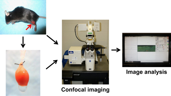
Figure 1. Schematic illustration of the confocal imaging of skeletal muscles and Langendorff perfused heart. The transgenic mouse expressing mt-cpYFP is anesthetized and the skeletal muscles on one of the hindlimbs are exposed for confocal imaging. The heart is then perfused in the Langendorff mode and confocal image is conducted. Image processing and data analysis used the confocal software and custom-developed program.
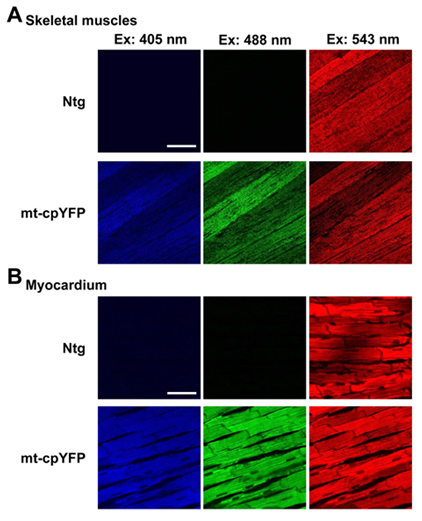
Figure 2. Confocal imaging of skeletal muscle fibers in vivo and myocardium in perfused heart. A. Representative images showing the skeletal muscles of nontransgenic (Ntg) and mt-cpYFP transgenic (mt-cpYFP) mouse loaded with mitochondrial membrane potential indicator, TMRM. B. Representative images showing the myocardium of Ntg and mt-cpYFP mice in Langendorff perfused heart. Ex: Excitation wavelength. mt-cpYFP is excited at 405 and 488 nm with emissions collected at 505-530 nm (blue) and 505-530 nm (green), respectively. TMRM is excited at 543 nm with emission collected at >560 nm (red). Scale bars = 50 μm.
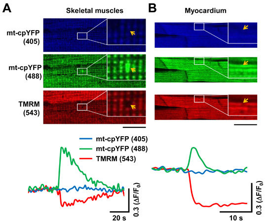
Figure 3. Single mitochondrial superoxide flashes detected in skeletal muscles in vivo and in perfused heart. A. Representative images (upper panel) and traces of time-dependent fluorescence change (mt-cpYFP at 405 and 488 nm excitation and TMRM at 543 nm excitation, lower panel) showing a single mitochondrial superoxide flash (highlighted by orange arrows in the enlarged portion of the images) in skeletal muscle fibers. Note the increased mt-cpYFP signal (at 488 nm excitation) is accompanied by decreased TMRM signal. B. Representative images (upper panel) and traces of time-dependent fluorescence change (lower panel) during a single mitochondrial superoxide flash in perfused myocardium. Scale bars = 50 μm. Click here to view larger figure.
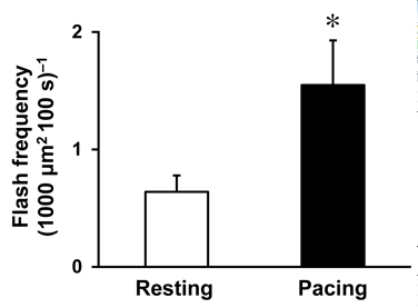
Figure 4. Increased superoxide flash frequency in perfused heart by electrical stimulation. The heart was electrically stimulated (Pacing, 4 V and 2 Hz) for 2 min. Data are mean ± SEM, n = 8 cells. *: P < 0.05 versus Resting. Click here to view larger figure.
Discussion
Imaging single mitochondrial events in live animal or perfused organs has significant advantage over traditional methods for mitochondrial function evaluation17,19,21,22,24,25. The technique described here can achieve real-time in situ determination of mitochondrial function in a real physiological condition at the subcellular resolution. This is particularly useful, when combined with other measurements, to systemically study the role of mitochondria in the normal function of a particular organ or cell type in vivo. This is a complicated technique that combines confocal microscopy and sophisticated perfusion system with the control of various parameters. Nevertheless, this technique has been used to link whole body glucose metabolism with muscle mitochondrial function17,19,20 and are currently employed by us to study the mitochondrial dysfunction in heart disease.
The Langendorff heart perfusion system is compatible to cardiac function evaluation through monitoring the left ventricle pressure and imaging intracellular Ca2+ transients during each beat. Alternatively, other fluorescent indicators such as MitoSOX or DCF can be perfused to facilitate in situ imaging of steady-state ROS levels in the myocardium. Langendorff perfused heart has been widely used to evaluate the response of heart to physiological (e.g. β-adrenergic stimulation by dobutamine) or pathological (e.g. ischemia reperfusion) perturbations26-28. Such treatments can be incorporated into the confocal imaging system to assess single mitochondrial function in response to stresses.
Limitations of this method include the selection of tissues/organs that are feasible for live animal imaging. Superficial tissues such as skeletal muscles, subcutaneous structures and superficial nerves are ideal for confocal imaging in live animal. Internal organs are technically inappropriate for live animal imaging due to the extensive damage to the mouse while exposing the organ. However, internal organs can be set up for organ perfusion/culture, such as Langendorff perfused heart or brain slice culture, if a physiological mimetic environment can be maintained. In addition, confocal microscope has an effective imaging depth of 30-50 μm, which allows the imaging of a few layers of cells beneath the organ surface. It is possible that this system can be combined with a multi-photon microscope, which has a much higher penetration depth (e.g. up to 1 mm) and has been used to assess mitochondrial function in the intact heart29. Motion artifact is another critical issue that should be considered. The mitochondria in skeletal muscles and cardiac myocytes showed minimal movement during the scan. Further, heartbeat-induced motion artifact can be suppressed or eliminated by perfusion with blebbistatin and application of gentle pressure on the heart. In other cell types, such as neurons and fibroblasts, the mitochondria may undergo constant and dramatic movements. During imaging processing and analysis, any signal changes due to motion artifact should be carefully identified and excluded.
Additional challenges of this technique include maintaining the appropriate condition throughout the experiment, positive controls and data interpretation. For skeletal muscle imaging in anesthetized mice, the exposed muscle surface should be immersed in physiological buffers at all time. Vital signs of the mouse such as breathing should be monitored throughout the experiment. For perfused heart studies, appropriate oxygenation, temperature control and substrate supply are required. Positive controls are recommended to verify the condition and responsiveness of the tissues. To do so, mice can be injected with insulin or glucose and perfused hearts can be electrically stimulated or perfused with isoproterenol. Increased superoxide flashes should be observed after these treatments (Figure 4)19,20. In addition, the time-lapse images are taken when the motion artifact is suppressed through inhibiting heartbeat, which produces sub-physiological conditions due to decreased work load. Thus, the perfused heart experiment should be considered as a close mimetic of the in vivo situation.
Finally, the technique was developed using a Zeiss confocal (LSM 510). Other confocal system (e.g. Nikon, Olympus or Leica) can be used if the system meets the following requirements: (1) has time-lapse frame scan mode, (2) has a sampling rate of 1 sec/frame, and (3) can reach single mitochondrion resolution (e.g. 1 μm x 2 μm). The software of a confocal system usually allows simple image processing and analysis, such as defining the ROIs and fluorescence intensity output with time labels. Alternatively, Image J software can be used for both image analysis and flash parameter calculation.
In summary, the imaging of single mitochondrial events such as superoxide flashes in skeletal muscles in vivo or in perfused heart is a unique approach for real-time determination of mitochondrial function. This technique is a significant advancement over the traditional in vitro measurements and will provide a more accurate evaluation of mitochondrial function and its relationship with cellular processes/functions under real physiological conditions.
Disclosures
The authors declare that they have no competing financial interests.
Acknowledgements
The authors would like to thank Drs. Heping Cheng, Huiliang Zhang and Stephen Kolwicz for their helpful comments and technical support in developing this method. This study was supported by NIH grants and the Scientist Development Grant from American Heart Association to WW.
Materials
| Name | Company | Catalog Number | Comments |
|---|---|---|---|
| REAGENTS | |||
| Blebbistatin | Toronto Research Chemicals | B592500 | |
| CaCl2 | Acros Organics | AC34961-5000 | |
| EDTA | Fisher Scientific | BP120-500 | |
| D-Glucose | Sigma-Aldrich | G8270-1 | |
| HEPES | Sigma-Aldrich | H7006-500 | |
| KCl | Sigma-Aldrich | P9541-1 | |
| MgCl2•6H2O | Fisher Scientific | BP214-500 | |
| MgSO4•7H2O | Sigma-Aldrich | M1880-1 | |
| NaCl | Fisher Scientific | BP358-212 | |
| NaH2PO4 | Sigma-Aldrich | S8282-500 | |
| NaHCO3 | Sigma-Aldrich | S6014-1 | |
| Pyruvate | Sigma-Aldrich | P2256-25 | |
| TMRM | Invitrogen | T-668 | |
| EQUIPMENT | |||
| Confocal Line Scanning Microscope (LSM 510 Meta, Zeiss), software version 4.2 SP1 including "Physiological Analysis" module. |
References
- Brookes, P. S., Yoon, Y., Robotham, J. L., Anders, M. W., Calcium Sheu, S. S. Calcium, ATP, and ROS: a mitochondrial love-hate triangle. Am. J. Physiol. Cell Physiol. 287, 817-833 (2004).
- Scheffler, I. E. Mitochondria. , 2nd, J. Wiley and Sons, Inc. (2008).
- Rosca, M. G., Hoppel, C. L. Mitochondria in heart failure. Cardiovasc. Res. 88, 40-50 (2010).
- Griffiths, E. J. Mitochondria and heart disease. Adv. Exp. Med. Biol. 942, 249-267 (2012).
- Winklhofer, K. F., Haass, C. Mitochondrial dysfunction in Parkinson's disease. Biochim. Biophys. Acta. 1802, 29-44 (2010).
- Pieczenik, S. R., Neustadt, J. Mitochondrial dysfunction and molecular pathways of disease. Exp. Mol. Pathol. 83, 84-92 (2007).
- Szentesi, P., Zaremba, R., van Mechelen, W., Stienen, G. J. M. ATP utilization for calcium uptake and force production in different types of human skeletal muscle fibres. J. Physiol. 531, 393-403 (2001).
- Lemieux, H., Hoppel, C. L. Mitochondria in the human heart. J. Bioenerg. Biomembr. 41, 99-106 (2009).
- Chance, B., Williams, G. R. Respiratory enzymes in oxidative phosphorylation. III. The steady state. J. Biol. Chem. 217, 409-427 (1955).
- Lambert, A. J., Brand, M. D. Reactive oxygen species production by mitochondria. Methods Mol Biol. 554, 165-181 (2009).
- Brand, M. D., Nicholls, D. G. Assessing mitochondrial dysfunction in cells. Biochem. J. 435, 297-312 (2011).
- Robinson, K. M., et al. Selective fluorescent imaging of superoxide in vivo using ethidium-based probes. Proc. Natl. Acad. Sci. U.S.A. 103, 15038-15043 (2006).
- Dickinson, B. C., Srikun, D., Chang, C. J. Mitochondrial-targeted fluorescent probes for reactive oxygen species. Curr. Opin. Chem. Biol. 14, 50-56 (2010).
- Dooley, C. T., et al. Imaging dynamic redox changes in mammalian cells with green fluorescent protein indicators. J. Biol. Chem. 279, 22284-22293 (2004).
- Hanson, G. T., et al. Investigating mitochondrial redox potential with redox-sensitive green fluorescent protein indicators. J. Biol. Chem. 279, 13044-13053 (2004).
- Belousov, V. V., et al. Genetically encoded fluorescent indicator for intracellular hydrogen peroxide. Nat. Methods. 3, 281-286 (2006).
- Wang, W., et al. Superoxide Flashes in Single Mitochondria. Cell. 134, 279-290 (2008).
- Wei-Lapierre, L., et al. Respective Contribution of Mitochondrial Superoxide and pH to Mitochondria-targeted Circularly Permuted Yellow Fluorescent Protein (mt-cpYFP) Flash Activity. J. Biol. Chem. 288, 10567-10577 (2013).
- Fang, H., et al. Imaging superoxide flash and metabolism-coupled mitochondrial permeability transition in living animals. Cell Res. 21, 1295-1304 (2011).
- Wei, L., et al. Mitochondrial superoxide flashes: metabolic biomarkers of skeletal muscle activity and disease. FASEB J. 25, 3068-3078 (2011).
- Sheu, S. S., Wang, W., Cheng, H., Dirksen, R. T. Superoxide flashes: illuminating new insights into cardiac ischemia/reperfusion injury. Future Cardiol. 4, 551-554 (2008).
- Wei, L., Dirksen, R. T. Perspectives on: SGP symposium on mitochondrial physiology and medicine: mitochondrial superoxide flashes: from discovery to new controversies. J. Gen. Physiol. 139, 425-434 (2012).
- Johnson, I., Spence, M. T. Z. Ch. 12.2. The Molecular Probes Handbook, A Guide to Fluorescent Probes and Labeling Technologies. , Life Technologies Corp. 510-511 (2013).
- Wang, X., et al. Superoxide flashes: elemental events of mitochondrial ROS signaling in the heart. J. Mol. Cell Cardiol. 52, 940-948 (2012).
- Li, K., et al. Superoxide flashes reveal novel properties of mitochondrial reactive oxygen species excitability in cardiomyocytes. Biophys. J. 102, 1011-1021 (2012).
- Bell, R. M., Mocanu, M. M., Yellon, D. M. Retrograde heart perfusion: The Langendorff technique of isolated heart perfusion. J. Mol. Cell. Cardiol. 50, 940-950 (2011).
- Pasdois, P., et al. Effect of diazoxide on flavoprotein oxidation and reactive oxygen species generation during ischemia-reperfusion: a study on Langendorff-perfused rat hearts using optic fibers. Am. J. Physiol. 294, H2088-H2097 (2008).
- Granville, D. J., et al. Reduction of ischemia and reperfusion-induced myocardial damage by cytochrome P450 inhibitors. Proc. Natl. Acad. Sci. U.S.A. 101, 1321-1326 (2004).
- Davidson, S. M., Yellon, D. M., Murphy, M. P., Duchen, M. R. Slow calcium waves and redox changes precede mitochondrial permeability transition pore opening in the intact heart during hypoxia and reoxygenation. Cardiovasc. Res. 93, 445-453 (2012).
Reprints and Permissions
