Through a double treatment with colchicine, a plant-derived toxin that kills dividing cells, nerve-free Hydra vulgaris can be generated. These Hydra cannot feed or egest on their own. This paper describes an improved method for long-term maintenance of nerve free Hydra vulgaris in the laboratory.
Method Article
Generation and Long-term Maintenance of Nerve-free Hydra
In This Article
Summary
Abstract
The interstitial cell lineage of Hydra includes multipotent stem cells, and their derivatives: gland cells, nematocytes, germ cells, and nerve cells. The interstitial cells can be eliminated through two consecutive treatments with colchicine, a plant-derived toxin that kills dividing cells, thus erasing the potential for renewal of the differentiated cells that are derived from the interstitial stem cells. This allows for the generation of Hydra that lack nerve cells. A nerve-free polyp cannot open its mouth to feed, egest, or regulate osmotic pressure. Such animals, however, can survive and be cultured indefinitely in the laboratory if regularly force-fed and burped. The lack of nerve cells allows for studies of the role of the nervous system in regulating animal behavior and regeneration. Previously published protocols for nerve-free Hydra maintenance involve outdated techniques such as mouth-pipetting with hand-pulled micropipette tips to feed and clean the Hydra. Here, an improved protocol for maintenance of nerve-free Hydra is introduced. Fine-tipped forceps are used to force open the mouth and insert freshly killed Artemia. Following force-feeding, the body cavity of the animal is flushed with fresh medium using a syringe and hypodermic needle to remove undigested material, referred to here as "burping". This new method of force-feeding and burping nerve-free Hydra through the use of forceps and syringes eliminates the need for mouth-pipetting using hand-pulled micropipette tips. It thus makes the process safer and significantly more time efficient. To ensure that the nerve cells in the hypostome have been eliminated, immunohistochemistry using anti-tyrosine-tubulin is conducted.
Introduction
The nervous system of Hydra consists of a nerve net, with neurons associated with both epithelial tissue layers1. The nerve net is denser in the hypostome and peduncle and less dense in the body column2. The nerve cells originate from interstitial stem cells, which are multipotent stem cells that give rise to secretory cells, nematocytes, germ cells, and neurons1. It is possible to eliminate the interstitial cells of Hydra vulgaris through treatment with colchicine3,4, a plant-derived toxin that kills dividing cells. Although colchicine has been found to inhibit microtubule polymerization in other organisms, a previous study has shown that microtubules are present in Hydra throughout the entire treatment, suggesting that colchicine does not act this way in Hydra3. Another study suggests that colchicine does not bind efficiently to tubulin in some organisms, including Tetrahymena pyriformis, Zea mays, Chlamydomonas, and Schizosaccharomyces pombe, which may explain this difference5. The colchicine treatment induces phagocytosis of the interstitial cells by the endodermal epithelial cells3 and thus allows for the creation of animals that are lacking nerve cells, gland cells, and nematocytes. It is unclear why the interstitial cells are particularly susceptible to colchicine treatment. Given that both post-mitotic interstitial cells and the interstitial stem cell lineage are damaged and phagocytosed, Campbell concluded that colchicine was not directly affecting mitotic activity3. Notably, the colchicine treatment works well in Hydra vulgaris, but has been shown to not work as well in other species, such as Hydra oligactis6. A modified treatment with colchicine and hydroxyurea can be used to produce nerve-free Hydra viridis7. Nerve-free Hydra (also sometimes referred to as "epithelial Hydra"8) are therefore a useful tool for studying the roles of these specialized cell types from the interstitial cell lineage in tissue homeostasis and regeneration.
Hydra may be the only known example of an animal capable of living without a nervous system. Nerve-free Hydra serve as a particularly useful model for dissecting the role of the nerve net in regulating Hydra regeneration, homeostasis, and behavior. For example, the introduction of interstitial cells into nerve-free Hydra via grafting allowed for the characterization of nerve cell differentiation as highly region-specific9. Furthermore, because nerve-free Hydra can regenerate, they enable the investigation of alternative, nervous system-independent regeneration pathways. One such example is apical neurogenesis and head formation, which has been shown to depend on cnox-2 function in the nervous system in wildtype Hydra, but appears to be dispensable in nerve-free Hydra, suggesting that there may be an alternative head regeneration process10.
Nerve-free Hydra have also been used to study epithelial cell expression and regulation of neurogenic and neurotransmission genes after the loss of neurogenesis11. Nerve-free Hydra do not exhibit spontaneous contraction bursts12, indicating that these bursts are regulated by the nervous system. Nerve-free Hydra do, however, contract in response to pinching the body column with forceps, suggesting that contraction in response to mechanical stimuli is mediated by coupling through gap junctions in epithelial cells, while spontaneous contractile behavior is mediated by coupling through gap junctions in nerve cells13.
Nerve-free Hydra do not open their mouths when presented with food or reduced glutathione3, suggesting that sensory neurons are necessary to detect the presence of food and signal the mouth to open. In addition, the nerve net seems to play a role in sensing osmotic pressure, because nerve-free animals are unable to autonomously regulate their internal hydrostatic pressure through mouth opening, causing their characteristic balloon-like appearance3,4 (Figure 1B). Regulation of hydrostatic pressure in nerve-free Hydra by frequent manual deflation led to a loss of some abnormal morphology in the hypostome and body column. However, chronic deflation led to interference with growth, elongation, budding, and tissue organization8.
Although nerve-free Hydra are unable to feed and egest on their own, it is possible to maintain them indefinitely in the laboratory by manually force-feeding and burping each animal. Previous publications have described methods of force-feeding and burping nerve-free Hydra, however these protocols involved the use of micropipette tips that must be hand pulled carefully to the appropriate size as well as use of a mouthpiece connected to the pipette by tubing14. Here, a simpler, safer, and more time-efficient method of feeding and burping is described.
In addition, previous studies involved checking for the absence of nerve cells through dissociation of fixed animals into individual cells and examination of cell morphology3,4,15. Here, immunohistochemistry with a monoclonal antibody against the tyrosinated carboxyl-terminus of alpha-tubulin was used as a complimentary method to maceration to check for the depletion of neurons in the hypostome13,16. Previous studies have shown that neurons in the peduncle can also be visualized using this antibody13, however these neurons as well as those in the body column are more difficult to make out. While immunohistochemistry is sufficient to confirm the absence of nerve cells in the hypostome and does not require expertise on cell type morphology, it cannot be used to check for the absence of the interstitial stem cells and the other derivatives of these cells. Dissociation and cell morphology studies are more rigorous and can give a quantitative account of the numbers of each cell type remaining following each stage of the treatment.
Protocol
1. Double Colchicine Treatment
- Make a 0.4% colchicine (weight/volume) solution in Hydra medium3,4,17.
Caution: Colchicine is acutely toxic, fatal if swallowed, may cause genetic defects, and may cause eye damage. Handle the powder in a fume hood and wear full Personal Protective Equipment (PPE). - Incubate Hydra vulgaris (AEP strain was used here) that have been starved for 24 h in 0.4% colchicine at a ratio of 5 Hydra per mL of colchicine solution in a Petri dish for 8 h at room temperature in the dark.
Note: 5 Hydra/mL colchicine is a guideline for determining how much solution to use to avoid overcrowding the dish with Hydra. Use the same volume of solution for subsequent cleanings and solution changes. - Remove the colchicine solution and replace with clean Hydra medium without colchicine. Wash the Hydra 5 times by serial transfer with a glass Pasteur pipette into clean Hydra medium before transferring to 50 µg/mL rifampicin in Hydra medium. Keep the Hydra in an 18 °C incubator.
Note: Sufficient 1,000X rifampicin stock (50 mg/mL) in dimethyl sulfoxide (DMSO) for 1-2 weeks of treatment can be stored at 4 °C. The exact amount can be determined by the total volume of solution used each day. For example, if there are 2 dishes, each containing 10 mL of solution that needs to be changed twice daily, a total of 40 mL of solution will be used in one day, corresponding to 40 µL of 1,000X stock per day or 560 µL over a two week period. The stock is then diluted 1:1000 in Hydra medium upon use.
Caution: DMSO is a combustible liquid and readily penetrates skin, allowing other dissolved chemicals into the body. Handle the solvent in a fume hood and wear full PPE. Of note, DMSO freezes at 4 °C - Change the rifampicin-containing Hydra medium twice daily until the Hydra stop expelling cells into the medium, which is generally by 1 week post-treatment. At this point, the medium can be changed once daily. During the days following the treatment, the Hydra will completely lose their tentacles. Avoid having the Hydra in contact with one another, as they may fuse together and result in oddly shaped Hydra (Figure 2A).
- To prevent contact, avoid swirling the dish in a circular motion. Instead, gently agitate the dish in vertical and horizontal directions until the Hydra are spread apart. Inspect the dish upon placing the Hydra into the 18 °C incubator and adjust as necessary.
Note: For the days that the medium is changed twice, change the medium once in the morning and again in the afternoon/evening.
- To prevent contact, avoid swirling the dish in a circular motion. Instead, gently agitate the dish in vertical and horizontal directions until the Hydra are spread apart. Inspect the dish upon placing the Hydra into the 18 °C incubator and adjust as necessary.
- Once the tentacles begin to form, begin force-feeding and burping the Hydra 3 to 4 times a week (see Sections 2 and 3). About 8-9 days following colchicine treatment, the Hydra will begin to grow their tentacles back.
Note: Tentacle regrowth varies among individuals. Some may take longer than 8 - 9 days before showing signs of tentacle growth. Those that do not regrow their tentacles as well as oddly shaped (Figure 2B) animals or animals too small to be fed should be removed. These Hydra will likely not survive the second treatment. The Hydra may also bud. However, some of the buds may still have interstitial cells and be able to eat on their own.- Remove any buds that can eat without assistance and detach from the parent animal. Identify these animals by adding live Artemia to the feeding dish and observing whether or not the animals catch and eat the Artemia on their own. 1 or 2 Hydra may also be able to eat on their own. These should also be discarded.
Note: Nerve-free Hydra have a characteristic balloon-like morphology, and tentacles that are shorter and thinner than normal (due to loss of nematocytes) and can thus easily be distinguished from normal looking animals (Figure 1A, 1B).
- Remove any buds that can eat without assistance and detach from the parent animal. Identify these animals by adding live Artemia to the feeding dish and observing whether or not the animals catch and eat the Artemia on their own. 1 or 2 Hydra may also be able to eat on their own. These should also be discarded.
- Three weeks following the first colchicine treatment, repeat the colchicine treatment (steps 1.1 - 1.5). A second colchicine treatment is necessary to eliminate the remaining interstitial cells and nerve cells3.
2. Force-Feeding
- Add Artemia cysts to a narrow and tall glass container that tapers at the bottom. Fill the container with Artemia water (6.72 M NaCl). Avoid exceeding 1 g of cysts per 1 L of water for a higher hatch yield.
- Cover the top of the container with parafilm and insert a 10 mL serological pipette through the parafilm into the container. Provide aeration by attaching tubing to an aquarium air pump and fitting the tubing around the pipette. Make sure that the pipette tip reaches the bottom of the container and that no cysts are settled at the bottom. The Artemia will hatch after 48 h.
Note: There are many different ways to hatch Artemia that may work just as well.
- Cover the top of the container with parafilm and insert a 10 mL serological pipette through the parafilm into the container. Provide aeration by attaching tubing to an aquarium air pump and fitting the tubing around the pipette. Make sure that the pipette tip reaches the bottom of the container and that no cysts are settled at the bottom. The Artemia will hatch after 48 h.
- Strain the Artemia from the Artemia water in an Artemia net (available from aquarium supply companies) and wash them for about 20 s in DI water before placing them in a dish with Hydra medium. The salinity of Artemia water is too high for Hydra, so the Artemia must be washed before they are used.
- Under a dissecting microscope, euthanize the Artemia by lightly squeezing them with a pair of fine forceps. Avoid squeezing hard enough to expel the guts, as this will stick to the forceps and make feeding difficult. Lightly squeezing the Artemia before feeding it to the Hydra will help with the digestion process14. Place the freshly killed Artemia close to the Hydra in the dish to facilitate fast feeding.
Note: All subsequent steps are done under a dissecting microscope. - Use one pair of forceps to hold the Hydra by the peduncle. Continue holding the peduncle during the process of feeding the Hydra. Using a second pair of forceps, pinch the body column in order to make the Hydra contract.
- While holding the tips of the second pair of forceps together, tap at the center of the hypostome (the domed structure at the oral end of the animal). This sometimes causes the mouth to open. If the mouth does not open by tapping, puncture the mouth by inserting the forceps into the center of the hypostome and then slightly release the pressure on the tips of the forceps. This should stretch out the mouth opening made by the puncture.
Note: The Hydra may deflate and collapse when the mouth is opened (Figure 2C). If this occurs, the Artemia may still be inserted into the Hydra. If it is too difficult to do so, move on to the next Hydra and return to the original Hydra some time later and try again. The Hydra tend not to deflate as much during subsequent mouth openings. - Quickly pick up Artemia with the forceps and insert them into the Hydra's gastric cavity. Insert as many Artemia as possible until the gastric cavity is full and more Artemia cannot be inserted without damaging the Hydra, accidentally pulling out any Artemia inside, or preventing the mouth from closing. On average, this is 5 - 6 Artemia, however up to 12 have been fed to the larger animals.
- If the mouth begins to close, stretch open again with the forceps as described above; however, caution should be taken as any Artemia already inside the animal may begin to come out when the mouth is reopened.
- If the Hydra is too small for a whole Artemia, cut the Artemia using a scalpel and feed the Hydra smaller pieces. If the Artemia does not stay inside the Hydra, it may be necessary to press the forceps against the Artemia to keep it inside the Hydra until its mouth closes.
- Transfer fed Hydra with a glass Pasteur pipette, carefully to avoid accidental burping, to a dish with fresh 50 µg/mL rifampicin in Hydra medium. If an Artemia was expelled from the Hydra while transferring, reinsert it while in the new dish.
Note: A glass pipette is used to transfer the Hydra, as it has been found that Hydra stick less to glass compared to plastic. The length of the pipette does not matter, but using 5 inch pipettes may be somewhat easier.
3. Burping
- Burp the Hydra to remove undigested material any time between 8 and 20 h after feeding.
- Sand down the point of a 30G hypodermic needle with P320 or similar grit sandpaper until the tip is flat.
Note: A 27G hypodermic needle would also work, but the smaller tip of the higher gauge is preferable. - Attach the needle to a disposable 1 mL plastic syringe and fill the syringe with fresh 50 µg/mL rifampicin in Hydra medium.
Note: Burping a single Hydra takes about 0.05 mL to 0.1 mL of solution. A 1 mL syringe is preferable since controlling the force and volume of the solution coming out is more difficult with a larger volume syringe. - Under a dissecting microscope, hold the peduncle of the Hydra with fine point forceps and tap at the center of the hypostome with the needle. If the mouth does not open, insert forceps into the hypostome and open the mouth as described above for feeding.
Note: All subsequent steps are done under a dissecting microscope. - Use the syringe to flush, very gently to avoid blowing away the entire animal, the gastric cavity with the 50 µg/mL rifampicin solution until all debris has been expelled. The needle does not necessarily need to be inserted into the Hydra if the size of the Hydra does not permit. The needle may be held close to the mouth opening and pointed directly towards the mouth.
- Using a glass Pasteur pipette, transfer burped Hydra to a dish with fresh 50 µg/mL rifampicin in Hydra medium.
4. Quality Control Via Immunohistochemistry
NOTE: The following protocol is adapted from protocols by Shenk, M. A, et al.18, and Böttger, A19. All steps are done at room temperature (RT) unless otherwise noted. A nutator can be used for the incubation steps and may improve staining quality. However, if the Hydra get tangled up with one another, all steps can also be performed without.
- Prepare the blocking solution: 10% fetal bovine serum (FBS) and 1% DMSO in 1x phosphate-buffered saline (PBS). Store this solution at 4 °C until use (steps 4.6 and 4.11). The solution can be stored for 1 - 2 days.
Note: All solutions used in this protocol must be properly collected as hazardous waste. - Relax Hydra in 200 µL of 2% urethane in Hydra medium for 1 min in 1.7 mL microcentrifuge tubes. Use 5 Hydra per tube for each of the 4 conditions: nerve-free, untreated, untreated with no primary antibody, and untreated with no secondary antibody.
Note: Do not exceed 1 min. The timing here is critical as animals will be in bad shape after spending too much time in urethane. - Remove the urethane and fix the Hydra in 200 µL of 4% paraformaldehyde (PFA) in Hydra medium for 15 min.
- Wash the samples 3 times with 1x PBS for 10 min each.
Note: All washes with PBS and PBSTx are done with 500 µL of solution. - Permeabilize with 500 µL of 0.5% Triton X-100 in PBS for 15 min.
- Remove the 0.5% Triton X-100 and add 500 µL of the blocking solution. Block the samples for at least 1 h.
- Dilute the anti-tyrosine-tubulin antibody 1:200 in blocking solution.
Note: This concentration was determined experimentally. If the signal in the controls is found to be too weak, the concentration may need to be increased. If there is nonspecific binding, the concentration may need to be decreased. Pre-incubation of the antibody may also help reduce nonspecific binding. - Remove the blocking solution from the samples and add 200 µL of the primary antibody to the nerve-free Hydra, untreated controls, and untreated controls with no secondary. For the untreated controls with no primary, only add 200 µL of blocking solution without the antibody.
- Incubate the samples overnight (>12 h) at 4 °C.
Note: Alternatively, incubate 5 - 6 h at RT. - Remove the primary antibody and wash the samples extensively with 0.3% Triton X-100 in 1x PBS (PBSTx).
Note: Save the primary antibody and store at 4 °C, as it can be reused at least 2 - 3 times. - Dilute goat anti-mouse hP secondary antibody 1:500 in blocking solution. Add 200 µL to the nerve-free Hydra, untreated controls, and untreated controls with no primary. For the untreated controls with no secondary, only add 200 µL of blocking solution without the antibody.
Note: Alternatively, a fluorescent secondary antibody can be used, for which steps 4.13 - 4.17 are not needed. Using a fluorescent secondary saves time and is generally adequate, but the hP secondary provides increased sensitivity. The concentration of secondary was determined experimentally. If the signals in the controls are found to be too weak, the concentration may need to be increased. If there is nonspecific binding, the concentration may need to be decreased. - Incubate the samples overnight at 4 °C.
- Remove the secondary antibody and wash the samples extensively with PBSTx.
- Prepare 1x PBT: 0.2% bovine serum albumin (weight/volume), and 0.05% Tween20 in 1x PBS. Incubate the samples in 1x PBT for 30 min.
- Remove the 1x PBT and incubate the samples in 1:1,000 NHS-fluorescein and 1:10,000 H2O2 in 1x PBT for 15 min in the dark. From this step on, keep the samples in the dark as much as possible, as the fluorescent signal will decrease as they are exposed to light.
Note: NHS-fluorescein was prepared following a detailed FISH protocol by King, R. S. and Newmark, P. A.20 - Wash the samples 3 times with PBSTx quickly, then 2 times for 30 min.
- Leave the samples in PBSTx overnight at 4 °C to continue washing.
- Do a few more washes with 1x PBSTx. To visualize cell nuclei, carry out the following optional steps for DNA counterstaining using 4', 6-diamindino-2-phenylindole, dihydrochloride (DAPI). Otherwise, the samples may be imaged now.
- Dilute 5 mg/mL DAPI stock to a working concentration of 1:500 in PBSTx and add 200 µL to each tube. Incubate the samples for 30 min.
Caution: DAPI is a known carcinogen. Wear full PPE. - Remove the DAPI and wash the samples 2 - 3 times with PBSTx before imaging with fluorescence microscopy.
Results
Immediately following the initial 8 h colchicine treatment, all Hydra survive. Some of these Hydra will have only tentacle stubs remaining (Figure 3A), while others will have completely lost their tentacles (Figure 3B). Over the following 1 or 2 days, the tentacles will continue to shrink until all Hydra have lost their tentacles. About 1 week following the treatment, the Hydra will show signs of tentacle regrowth, with small stub like tentacles (Figure 3C) before they fully regenerate their tentacles (Figure 3D). Of the original Hydra, around 50-60% eventually regenerate their tentacles between 1-2 weeks after the treatment.
Following the second treatment, the recovery of the animals is similar to that after the first treatment. Thus, around 10% of the original number of animals that went into both treatments recover and grow to a size suitable for experimentation (Figure 1B). These Hydra have tentacles that appear thinner than those of untreated animals, have tissue which is more transparent, and will appear bloated due to the inability to open their mouths in order to relieve osmotic pressure (Figure 1A, 1B). Any animals that are not fed can survive for a few weeks, however, unfed animals will shrink in size and become increasingly difficult to feed.
Hydra that do recover following the second treatment can be maintained indefinitely. These animals are able to bud, thus sustaining and growing the population. Additionally, the population may be grown by cutting these animals and allowing them to regenerate4.
Immunohistochemistry confirms the loss of neurons in the hypostome following the second colchicine treatment. The dense nerve net in the hypostome can be visualized using an anti-tyrosine-tubulin antibody13,16 and shows distinct fibers radiating outward from the mouth and obvious cell bodies (Figure 4A). These fibers are absent in nerve-free Hydra that have been produced by double-treatment with colchicine (Figure 4B). Furthermore, co-staining with DAPI reveals a reduced number of cell nuclei in the nerve-free Hydra compared to the untreated controls due to the loss of cells of the interstitial cell lineage as a result of the colchicine treatments (Figure 4A, 4B).

Figure 1. Comparison of Nerve-free and Untreated Hydra.
(A) Untreated Hydra. (B) Nerve-free Hydra 22 days following the second colchicine treatment. Note the swollen body column and the thin tentacles in the nerve-free animal. Scale bars are 500 µm. Please click here to view a larger version of this figure.

Figure 2. Examples of Deformed Hydra.
(A) Two Hydra that have fused together following the first treatment. (B) An oddly shaped Hydra. (C) A Hydra that has deflated after having its mouth opened. Scale bars are 400 µm. Please click here to view a larger version of this figure.
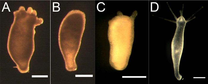
Figure 3. Representative Images of Hydra Following the First 8 h Colchicine Treatment.
(A) A Hydra with stubby tentacles immediately after treatment. (B) A Hydra that has completely lost its tentacles immediately after treatment. (C) A Hydra that is beginning to regrow its tentacles 8 days following the treatment. (D) A Hydra that has completely regrown its tentacles 13 days following the treatment. Scale bars are 400 µm. Please click here to view a larger version of this figure.
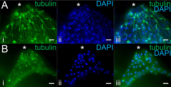
Figure 4. Loss of Nerve Cells in the Hypostome can be Confirmed by Immunohistochemistry.
(A) The hypostome of an untreated Hydra and (B) hypostome of a nerve-free Hydra approximately 2 weeks following the second colchicine treatment, labeled with (i) anti-tyrosine-tubulin antibody and (ii) DAPI. (iii) shows the overlay. The * indicates the location of the mouth. Maximum z projections were made from spinning disk confocal fluorescence z stacks taken with exposures of 500 ms (GFP) and 15 ms (DAPI). Brightness and contrast were adjusted to improve visibility. Scale bars are 20 µm. Please click here to view a larger version of this figure.
Discussion
Hydra interstitial cells can be eliminated through a double colchicine treatment3,4. In the days following the first treatment, it is critical to prevent contact between individual Hydra to avoid fusion of Hydra pieces into deformed Hydra. Also, animals that can eat unassisted following the first treatment must be removed as the second colchicine treatment may not be sufficient to eliminate the remaining interstitial cells from such animals. To maximize the survival rate of nerve-free Hydra after the second colchicine treatment, the animals should be fed as many Artemia as possible following recovery from the first treatment, with 1 - 2 days between feedings to recover and grow. Each colchicine treatment causes the Hydra to diminish significantly in size as cells are egested, so the larger the Hydra are prior to each treatment, the better the chances that the Hydra will recover to a size that can be fed and maintained.
Due to the low survival rate of Hydra after each treatment, it may be desirable to start with many Hydra. However, caution must be taken with starting treatment on too many Hydra at once. This will make feeding following the first treatment difficult and extremely time-consuming. Animals fed well after the first treatment recover to larger sizes and therefore have a higher survival rate after the second treatment. Thus, it may be more productive to start with fewer and focus on feeding better. Generally, starting with 50 - 100 animals is manageable for 1 - 2 people. It is also best to begin with the largest Hydra possible, as these will better survive the two treatments.
The method of force-feeding and burping Hydra that are lacking nerve cells described here is safer, simpler, and more time-efficient than methods described in the past3,4,14. The use of commercially available forceps and syringes eliminate the need for hand pulled micropipette tips, which are time consuming and difficult to make. The use of these tools also avoids the need to mouth-pipette. Force-feeding using forceps may be difficult at first but given enough time and practice, it becomes quite easy and efficient. One should first practice force-feeding and burping normal Hydra to get a feel for how much force to use with the tools and how to manipulate Hydra. Various forceps and needle sizes were tested to find the optimal tools for force-feeding and burping.
The use of antibody staining as described here is only adequate to check for the absence of nerve cells in the hypostome. To ensure that all interstitial cells have been eliminated, animals may be macerated and the types of cells present can be examined15.
Disclosures
The authors have nothing to disclose.
Acknowledgements
The authors thank Dr. Dick Campbell (UC Irvine) for discussions about the original protocol for generating and maintaining nerve-free animals, Ms. Rui Wang for help with adapting the syringe and needle technique, and Ms. Danielle Hagstrom and Dr. Rob Steele (UC Irvine) for comments on the manuscript. This work was supported by the RCSA and NSF grant CMMI-1463572.
Materials
| Name | Company | Catalog Number | Comments |
|---|---|---|---|
| Colchicine | Acros Organics | 227120010 | |
| 1 mL Syringe | BD | 301025 | |
| Brine Shrimp Eggs | Brine Shrimp Direct | N/A | Can be purchased locally |
| Brine Shrimp Hatchery Dish | Brine Shrimp Direct | N/A | |
| 60 mm x 15 mm Petri Dish | Celltreat | 229663 | |
| 30 G x 3/4" Hypodermic Needle | Covidien | 1188830340 | A 27G needle may also be used |
| 2 x Fine-tip Tweezers | Dumont | 0109-5-PO | |
| Rifampicin | EMD Millipore | 557303 | |
| Goat anti-mouse lgG, Pab (HRP Conjugate) | Enzo | ADI-SAB-100-J | |
| Bovine Serum Albumin (BSA) | Fisher | BP9703-100 | |
| Scalpel | Fisher | 08-920A | |
| Fetal Bovine Serum (FBS) | Gibco | 10437028 | |
| DAPI (4',6-Diamidino-2-Phenylindole, Dihydrochloride) | Invitrogen | D1306 | |
| PBS Tablets | MP Bio | 2810305 | |
| Monoclonal Anti-Tubulin, Tyrosine | Sigma | T9028 | |
| Dimethyl Sulfoxide (DMSO) | Sigma | D2650 | |
| Hydrogen Peroxide | Sigma | 216763 | |
| Paraformaldehyde (PFA) | Sigma | P6148 | |
| Triton X - 100 | Sigma | T9284 | |
| Tween 20 | Sigma | P1379 | |
| Urethane | Sigma | U2500 | |
| Air Pump | Tetra | 77846-00 | |
| Glass Pasteur Pipette | VWR | 53283-916 | Length of the pipet does not matter |
| 15 mL Tube | VWR | 89039-670 | |
| P320 Sandpaper | 3M | IBGABBV00397 | Can be purchased at local home improvement store |
References
- Bode, H. R. The interstitial cell lineage of Hydra: a stem cell system that arose early in evolution. J. Cell. Sci. 109, 1155-1164 (1996).
- Hager, G., David, C. N. Pattern of differentiated nerve cells in hydra is determined by precursor migration. Development. 124, 569-576 (1997).
- Campbell, R. D. Elimination of Hydra interstitial and nerve cells by means of colchicine. J. Cell. Sci. 21, 1-13 (1976).
- Marcum, B. A., Campbell, R. D. Development of Hydra lacking nerve and interstitial cells. J. Cell. Sci. 29, 17-33 (1978).
- Burns, R. G. 3H-colchicine binding: Failure to detect any binding to soluble proteins from various lower organisms. Exp. Cell. Res. 81, 285-292 (1973).
- Lee, H. -T., Campbell, R. D. Development and Behavior of an Intergeneric Chimera of Hydra (Pelmatohydra oligactis Interstitial Cells Hydra attenuata Epithelial Cells). Biol. Bull. 157 (2), 288-296 (1979).
- Novak, P. Preparing Hydra viridis with Nerve Cells and No Interstitial Cells, or with Neither of These Cell Types. Hydra: Research Methods. , Plenum Press. New York. 295-297 (1983).
- Wanek, N., Marcum, B. A., Lee, H. -T., Chow, M., Campbell, R. D. Effect of hydrostatic pressure on morphogenesis in nerve-free Hydra. J. Exp. Zool. 211, 275-280 (1980).
- Minobe, S., Koizumi, O., Sugiyama, T. Nerve cell differentiation in nerve-free tissue of epithelial Hydra from precursor cells introduced by grafting. Dev. Biol. 172 (1), 170-181 (1995).
- Miljkovic-Licina, M., Chera, S., Ghila, L., Galliot, B. Head regeneration in wild-type hydra requires de novo neurogenesis. Development. 134 (6), 1191-1201 (2007).
- Wenger, Y., Buzgariu, W., Galliot, B. Loss of neurogenesis in Hydra leads to compensatory regulation of neurogenic and neurotransmission genes in epithelial cells. Phil. Trans. R. Soc. B. 371 (20150040), (2015).
- Campbell, R. D., Josephson, R. K., Schwab, W. E., Rushforth, N. B. Excitability of nerve-free Hydra. Nature. 262, 388-390 (1976).
- Takaku, Y., Hwang, J. S., et al. Innexin gap junctions in nerve cells coordinate spontaneous contractile behavior in Hydra polyps. Sci. Rep. 4 (3573), (2014).
- Marcum, B. A. Culturing Epithelial Hydra. Hydra: Research Methods. , Plenum Press. New York. 287-290 (1983).
- David, C. N. A quantitative method for maceration of Hydra tissue. Wilhelm Roux Arch. Entwickl. Mech. Org. 171, 259-268 (1973).
- Gröger, H., Schmid, V. Larval development in Cnidaria: A connection to Bilateria. Genesis. 29 (3), 110-114 (2001).
- Lenhoff, H. M. Water, Culture Solutions, and Buffers. Hydra: Research Methods. , Plenum Press. New York. 29-34 (1983).
- Shenk, M. A., Bode, H. R., Steele, R. E. Expression of Cnox-2, a HOM/HOX homeobox gene in hydra, is correlated with axial pattern formation. Development. 117, 657-667 (1993).
- Böttger, A., et al. Horizontal Gene Transfer Contributed to the Evolution of Extracellular Surface Structures: The Freshwater Polyp Hydra Is Covered by a Complex Fibrous Cuticle Containing Glycosaminoglycans and Proteins of the PPOD and SWT (Sweet Tooth) Families. PLoS One. 7 (12), (2012).
- King, R. S., Newmark, P. A. In situ hybridization protocol for enhanced detection of gene expression in the planarian Schmidtea mediterranea. BMC Dev. Biol. 13 (8), (2013).
Reprints and Permissions
