7,393 Views
•
12:21 min
•
October 31, 2017
DOI:
The overall goal of this procedure is to fabricate and surface-modify polylactic acid, co-glycolic acid, electrospun fibers, with varying amounts of the antiviral protein Griffithsin. This method can help answer key questions in the drug delivery field, such as how to formulate and characterize new delivery vehicles that can prevent sexually transmitted viral infections, such as HIV-1. The main advantage of the technique is that the antiviral activity of Griffithsin is maximized by using polymeric-electrospun fibers as a stationary scaffold to highly localized Griffithsin on the fiber surface.
The implications of this technique extend toward therapy of other viral and bacteria infections, because of Griffithsin’s potent prophylactic and therapeutic potential against a variety of infection types. Though this method can provide insight into preventing HIV-1 infection, it can also be applied to other systems, such as other viral or bacterial infections in which Griffithsin has demonstrated potency. To begin this procedure, weigh 720 milligrams of 50/50 PLGA into a 10 milliliter scintillation vile.
Using a serological glass pipette, add 3.0 milliliters of HFIP. Cover the vile with plastic film. Then, measure and record the vile mass.
Incubate the polymer suspension at 37 degrees Celsius overnight to ensure complete dissolution of the polymer. After this, prepare the electrospinning apparatus as outlined in the text protocol. Use a three milliliter syringe to aspirate the polymer solution.
Next, connect a blunt 18 gauge 0.5 inch needle tip to the syringe. Dispense the the excess solution to remove empty head space in the needle. Place the syringe on a syringe pump, and set the instrument flow rate to 2.0 milliliters per hour.
Connect the power source to the syringe needle. Electrospin the polymer solution using a voltage of positive 27 kilovolts. After the entire solution is electrospun, turn off the power source.
Allow the mandril to spin for an additional 30 minutes, to fully evaporate the solvent. Then, turn off the rotating mandril collector. Using a razor blade, gently peel the fiber from the mandril.
Collect the electrospun PLGA fiber into a labeled petri dish. Place the dish in a desiccator overnight to remove any residual solvent. To begin, prepare the PBS and MES solutions as outlined in the text protocol.
Next, remove the EDC and NHS from the freezer. Let them rest to equilibrate to room temperature. After this, weigh four milligrams of EDC into a 1.5 milliliter micro-centrifuge tube.
Weigh six milligrams of NHS into a different micro-centrifuge tube. Add one milliliter of MES buffer to each tube. Vortex both tubes vigorously to ensure the reagents are fully dissolved.
Then, weigh 70 milligrams of hydroxylamine into a 50 milliliter conical centrifuge tube. Add 20 milliliters of PBS, and vortex. Mass out an appropriate amount of PLGA fiber into a 15 milliliter conical centrifuge tube.
Add eight milliliters of MES buffer to the fiber. Next, add one milliliter of both the EDC and NHS solutions. Use plastic film to seal the 15 milliliter conical centrifuge tube.
Place the tube on a rotator to gently invert the solution for 15 minutes at room temperature. After this, add 14 microliters of beta-mercaptoethanol to quench the reaction. Invert the tube several times to ensure complete mixing.
Discard the supernatant, and rinse the PLGA fiber twice using 10 milliliters of PBS for each wash. Add an appropriate amount of GRFT stock solution to the tube, as outlined in the text protocol. Then, add enough PBS to bring the final volume to eight milliliters.
Close and invert the tube to ensure thorough mixing. Seal the tube with plastic film. Place the tube on the rotator to gently invert the solution for two hours.
After this, quench the reaction by adding two milliliters of the prepared hydroxylamine solution, and vortexing for 30 seconds. Discard the supernatant. Rinse the surface modified PLGA fiber twice, using 10 milliliters ultra pure water, and vortexing for each wash.
Next, transfer the washed fiber to a petri dish. Place the petri dish inside of a desiccator, until the fiber is completely dry. Then store the petri dish at four degrees Celsius.
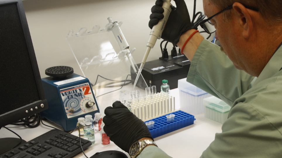
To begin, set out three 1.5 milliliter micro-centrifuge tubes. Mass out two milligrams of fiber into each. Then, add one milliliter of DMSO to each tube.
Vortex for one minute at room temperature to completely dissolve the fiber. After this, dilute a 10 microliter aliquot at least 100 fold, in TE buffer. Store at minus 20 degrees Celsius, until loading characterization with Eliza.
Weigh out between 5 and 10 milligrams of surface modified fiber, and transfer it to a micro-centrifuge tube. Record the fiber mass placed in each tube. Next, add one milliliter of a solution that mimics the physiological environment of each sample.
Incubate the samples on a rotating shaker at 200 RPM and 37 degrees Celsius for one hour. After this, remove approximately one milliliter of the solution containing the desorbed GRFT and aliquot it into cluster tubes. Store at minus 20 degrees Celsius until ready to perform protein quantification.
Then transfer the sample to a new micro-centrifuge tube. Add one milliliter of fresh buffer solution and incubate until the next time point, as outlined in the text protocol. In this study, the conditions for fabricating PLGA electrospun fibers are examined.
SEM analysis of blank, 0.05 nanomole, 0.5 nanomole, and 5 nanomole GRFT fibers indicates that GRFT modification has no effect on fiber morphology. All of the formations are seen to have similar average diameters, around 1.9 microns, demonstrating the consistency of the modified fabrication process across batches. The amount of GRFT conjugated in the electrospun fibers is then determined.
The 5 nanomole modification contains 373 nanograms of GRFT, while the 0.5 and 0.05 modifications contains 165 and 42, respectively. This demonstrates that fibers conjugated with higher theoretical surface modification density result in more GRFT conjugated to the fiber. However, an inverse correlation can be seen in the resulting conjugation efficiency.
Next, the amount of GRFT covalently conjugated to the fiber is assessed. Within the first four hours, 113 nanograms, 25 nanograms, and 10 nanograms of GRFT per milligram of electrospun fiber is detected in the 5 nanomole, 0.5 nanomole, and 0.05 nanomole theoretical modification concentrations, respectively. After four hours, negligible GRFT is detected in the release elute for all three formulations.
This data indicates that the majority of GRFT is covalently bound to the electrospun fibers and that the surface absorbed GRFT is released within the first four hours. Once mastered, this technique can be done in approximately four days. Electrospinning the polymer fiber scaffold, requires two days to prepare the polymer solution and to electrospin.
Whereas surface modification, subsequent drying and fiber characterization required at least two additional days. While attempting this procedure, it’s important to remember to maintain the proper ratios of reagents to ensure optimal modification. Furthermore, it’s also important to verify that the electospinning parameters are correct to ensure appropriate fiber morphology.
After its development, this technique paved the way for researchers in the field of drug delivery to fabricate electrospun fibers, that not only encapsulate antiviral agents within the fiber, but have a capacity to conjugate active agents through the fiber surface. After watching this video, you should have a good understanding of how to electrospin polymeric fiber, perform surface modification of fiber with the anti-viral protein Griffithsin, and qualitative Griffithsin conjugated to the fiber. Don’t forget that working with reagents such as HFIP, beta-mercaptoethanol, EDC and NHS can be extremely hazardous.
Precautions such as proper application of PPE, and the use of chemical fume hoods, should always be taken while performing this procedure.
Este manuscrito descreve o procedimento para fabricar e caracterizar as fibras de electrospun de Griffithsin-modificado poli (ácido lático-co-glicólico) que demonstram potente atividade antiviral e adesiva contra infecção por vírus da imunodeficiência humana tipo 1 em vitro. Métodos utilizados para sintetizar, modificar a superfície e caracterizar a morfologia resultante, conjugação, e dessorção de Griffithsin superfície-modificado com fibras são descritos.
Read Article
Cite this Article
Vuong, H. R., Tyo, K. M., Steinbach-Rankins, J. M. Fabrication and Characterization of Griffithsin-modified Fiber Scaffolds for Prevention of Sexually Transmitted Infections. J. Vis. Exp. (128), e56492, doi:10.3791/56492 (2017).
Copy