7,161 Views
•
08:56 min
•
November 15, 2017
DOI:
The overall goal of this procedure is to fabricate a small, ready to use cassette that can be applied for visual detection of multiple nucleic acids in a single, easy to operate test. This method can help answer key questions in the multiplex nucleic acid detection field, such as genetically modified organisms, or GMO detection. To clean the capillaries, don a lab uniform, safety goggles, and chemical protective gloves.
Place capillaries into a beaker. Clean the capillaries with 30 milliliters of acetone for five minutes to remove organic matter. And then, wash the capillaries with deionized water.
Next, prepare the piranha solution by pouring about ten milliliters of hydrogen peroxide into the beaker. Then, add 30 milliliters of sulfuric acid into the hydrogen peroxide with slow shaking to prevent overheating. Make sure that the solution completely covers the capillaries for at least 30 minutes.
Then, wash away the piranha solution with a large amount of water. Wash the capillaries with ethanol and deionized water for five minutes each. Dry the capillaries in a drying oven.
Next, weigh PDMS base and gearing reagents with a ratio of one-to-one in a 50 milliliter tube. Typically, mix five grams of elastomer base with five grams of elastomer curing agent. Stir the mixture thoroughly for about five minutes with a glass rod.
Place the beaker with the PDMS in a vacuum bell jar for thirty minutes for degassing. Then, pour the PDMS slowly into the cylinder of the stainless steel mold. Allow the PDMS to cure for three hours in a drying oven at 60 degrees Celsius.
After curing the PDMS remove the mold by pulling out the mold with cylindar. Remove the PDMS from the cylinder by cutting the margin of the mold with a scalpel. Wash PDMS three times with ethanol and deionized water.
Then, dry the PDMS support with nitrogen. Treat the lower surface of the PDMS support to be hydrophobic by soaking the PDMS support in a super hydrophobic coat for one second. Then, dry it for ten minutes at room temperature.
Next, insert the cleaned four millimeter capillaries into the holes of the PDMS support, leaving zero point five millimeters of the capillaries outside the top surface of the PDMS support. Make sure the ends of the capillaries are on the same level. To treat the outer surface of the capillaries and the top surface of the PDMS support to be hydrophobic, add 15 microliters of the super hydrophobic coat on top of the surface of the PDMS support.
Air dry the super hydrophobic modified capillary array. Next, add one point six microliters of the primer fixing mixture of one set of primers to fill one corresponding capillary of the capillary array according to a pre-designed order. Anchor the array in a transparent well of a standard flat bottom 96-well plate and dry the array at 60 degrees Celsius for at least two hours.
To assemble the cassette, place a loading adapter onto the top of the anchored array. Make sure the dish of the adapter just covers the exposed parts of all ten capillaries. To prepare the LAMP reaction mix, add reagents to the mixture according to the text protocol and vortex the reaction mixture for five seconds after adding calcine.
Gently invert the tube twenty times after Bst polymerase is added. Trace DNA that exists in the environment can be eliminated by cleaning earlier with one mole per liter of hydrochloric acid before handing the LAMP mix. Aspirate 20 microliters of the LAMP reaction mixture with a standard 100 microliter tip.
Insert the tip into the inlet of the loading adapter to lock it. Gently inject the reaction mixture into the dish of adapter. The reaction mixture will quickly fill the dish, and then load the capillaries automatically through capillary force.
Remove the adapter with the lock tip and seal the well by a PCR compatible transparent sealing film. Then, incubate the capillary array in an incubator at 63 degrees Celsius for one hour. To acquire images of the fluorescence emission, use a UV flashlight to excite the dissociated calcine to emit fluorescence.
Capture images from the top of the capillary array by either a digital camera or a smart phone. When taking an image, ensure that the camera is zoomed in on the area of the capillaries as much as possible to get high quality, clear images. Next, open the image by image analysis software.
And then select image, mold, grayscale, yes and image, mold, 16-bit, yes. Select file, save as, format, TIFF, to convert the image to 16-bit TIFF format. To extract the value of fluorescence intensity, open the microarray analysis software.
Drag the 16-bit TIFF format image into the interface of the software, and then select the wavelength of 532 nanometers and a color of green to display the image. Then, create a new block to locate the fluorescence signal from the capillaries. Select tools, new blocks, and then put the number of columns and rows as one-to-one and two-to-five in the blocks and features interfaces separately.
Right click and select the features model. Then, adjust the location and diameter to fit the fluorescence area of the capillaries. Select analyze to extract the value of fluorescence intensity.
Finally, select file, save settings as, to save the block’s documents. Then, select, file, save results as to save the fluorescence intensity results. Shown here is the layout of the capillary array.
Capillaries one through eight, prefixed with LAMP primer sets. Capillaries nine and ten represent two, no primer controls. Shown here is the fluorescent photograph for the detection of genetically modified maize, MON863, where the green color represents the positive LAMP amplification.
Successful amplification, only presented in expected capillaries one, six, seven and eight, without contamination among blank capillaries. This fluorescent photograph is for the detection of the GMO mix. The green color presented the positive LAMP amplification.
Successful amplification only presented in expected capillaries one, four, five, six, seven and eight without contamination among blank capillaries. The fluorescent photograph shown here is for the detection of non-genetically modified maize, with the green color presenting the positive LAMP amplification. Successful amplification only presented in the expected capillary eight, and without contamination among blank capillaries.
Once mastered, this detection procedure can be done in one point five hours if it is performed properly. After watching this video, you should have a good understanding of how to fabricate capillary-based cassette to perform multiple nucleic acid detection.
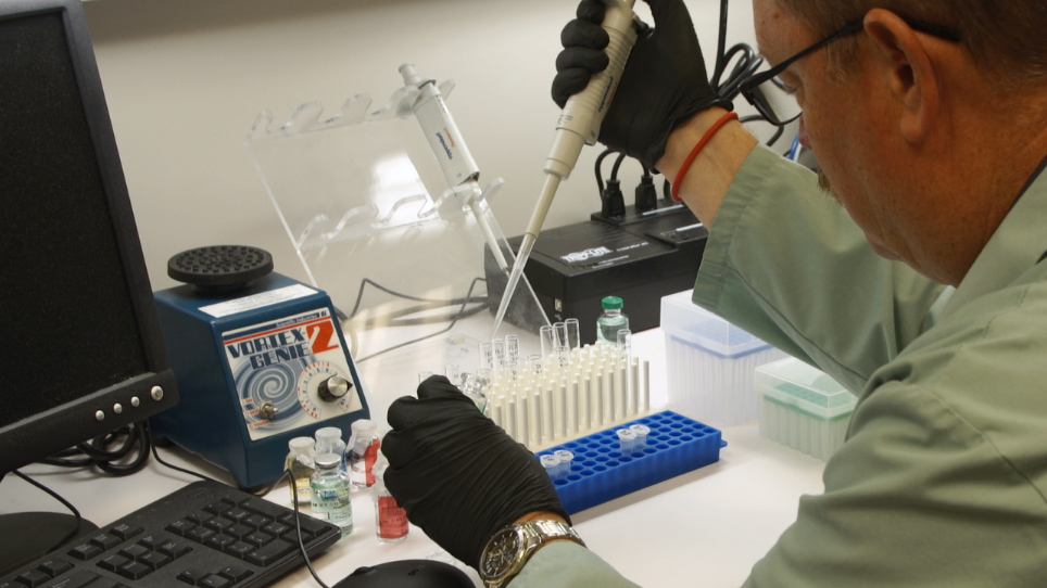
Este protocolo describe la fabricación de una cinta pequeña, lista para usar que se puede aplicar para detección visual de varios ácidos nucleicos en una prueba individual, que es fácil de operar. En este enfoque, una gama capilar fue utilizada para la detección multiplex y altamente eficiente de los objetivos de la OMG.
Read Article
Cite this Article
Chen, J., Shao, N., Hu, J., Li, R., Zhu, Y., Zhang, D., Guo, S., Hui, J., Liu, P., Yang, L., Tao, S. Visual Detection of Multiple Nucleic Acids in a Capillary Array. J. Vis. Exp. (129), e56597, doi:10.3791/56597 (2017).
Copy