Journal
/
/
Синтез Aptamer-PEI-g-PEG Модифицированные золотые наночастицы загружены доксорубицином для целевой доставки лекарств
JoVE Journal
Chemistry
A subscription to JoVE is required to view this content.  Sign in or start your free trial.
JoVE Journal Chemistry
Synthesis of Aptamer-PEI-g-PEG Modified Gold Nanoparticles Loaded with Doxorubicin for Targeted Drug Delivery

Синтез Aptamer-PEI-g-PEG Модифицированные золотые наночастицы загружены доксорубицином для целевой доставки лекарств

5,699 Views

09:09 min

June 23, 2020

DOI:

09:09 min
June 23, 2020

6 Views
, , , , , ,

Transcript

Automatically generated

Because of the drug resistance and toxicity, the use of doxorubicin is limit in the clinic. This protocol provides a degradable carrier for keeping the release of this therapeutic agent. The advantage of this method is that the synthesized carrier can be used to deliver the drug directly to cancer cells, inducing cell death in the cancer cells only.

For CT-PEG synthesis, first add 1.46 grams of succinic anhydride and 209 milligrams of 4-dimethylaminopyridine to a 100 milliliter round bottom flask. Next, add 15 milliliters of anhydrous tetrahydrofuran to the flask and fit a glass stopper into the flask, before incubating the reaction for 30 minutes at zero degrees Celsius. During the incubation, add 4.208 grams of PEG, 1.8 milliliters of triethylamine and 15 milliliters of anhydrous tetrahydrofuran to a new flask and close the flask with a glass stopper.

At the end of the incubation, use a syringe under nitrogen atmosphere to slowly transfer the second solution to the first flask. Stir the new solution for two hours at zero degrees Celsius, before continuing the reaction at room temperature overnight. The next morning, use a rotary evaporator at 40 degrees Celsius and 0.1 megapascals to concentrate the reaction solution and to remove the tetrahydrofuran solvent.

After an hour, dissolve the reaction solution in 15 milliliters of 1.325 gram per milliliter of dichloromethane at room temperature, before adding 15 milliliters of cold diethyl ether to obtain the PEG diacid precipitation product. Then remove the solvent via filter paper and dry the precipitate under vacuum for 48 hours at room temperature. For PEI-g-PEG copolymer synthesis, add 305.47 milligrams of the CT-PEG precipitate and five milliliters of DMSO to a new flask and stir the reaction at room temperature until the CT-PEG is fully dissolved.

Next, dissolve 49.46 milligrams of EDC in five milliliters of DMSO and add the solution to the flask. Stir the reaction for 30 minutes at room temperature, before dissolving 29.69 milligrams of N-Hydroxysuccinimide and five milliliters of DMSO and adding the resulting solution to the flask. Continue to stir the reaction for three hours at room temperature.

Near the end of the incubation, dissolve 28.6 microliters of PEI into 10 milliliters of DMSO and add the solution drop wise to the flask. Stir the reaction at room temperature for at least three days. At the end of the incubation, transfer the reacted solution to a dialysis bag with a 1000 molecular weight cutoff and place the bag into a one liter beaker containing 500 milliliters of ultra pure water as the dialysate.

At the end of the dialysis, transfer the solution to a dialysis bag with a 10, 000 molecular weight cutoff and placed the bag into a one liter beaker with 500 milliliters of ultra pure water as the dialysate. At the end of the second dialysis, use a rotary evaporator at 40 degrees Celsius and 0.1 megapascals to concentrate the solution and freeze dry the sample to obtain the PEI-g-PEG powder. For the coating of gold nanoparticles with the PEI-g-PEG product, dissolve five milligrams of the prepared PEI-g-PEG in five milliliters of ultra pure water in a new flask and fit the flask with a glass stopper.

Add 100 milliliters of 0.3 millimolar auric chloride solution to the flask and stir the solution for three hours at room temperature. The colors should change immediately from light yellow to dark yellow. At the end of the incubation, add one milliliter of one milligram per milliliter of sodium borohydride solution to the flask.

The color should change immediately from dark yellow to golden yellow, and stir the solution for an additional three hours at room temperature. Then dialyze the reaction product with a 1000 molecular weight cutoff dialysis bag for three days as demonstrated to obtain the PEI-g-PEG coated gold nanoparticle solution. For grafting of doxorubicin to the PEI-g-PEG coated gold nanoparticles.

Add one milliliter of 2.2 milligram per milliliter doxorubicin solution and 20 milliliters of the PEI-g-PEG coated gold nanoparticle solution to a new flask and fit the flask with a glass stopper. Next, dissolve 0.727 milligrams of EDC in one milliliter of ultra pure water and add the EDC solution to the flask. Dissolve 0.437 milligrams of N-Hydroxysuccinimide in one milliliter of ultra pure water, and add the N-Hydroxysuccinimide solution to the flask for a one hour incubation with stirring at room temperature.

At the end of the incubation, dialyze the reaction product in a 1000 molecular weight cutoff dialysis bag for three days as demonstrated to obtain the doxorubicin g-PEI-g-PEG coated gold nanoparticle solution. For grafting of the AS1411 aptamer to the DOX-g-PEI-g-PEG. Add 20 milliliters of the doxorubicin DOX-g-PEI-g-PEG coated gold nanoparticle solution to an approximately four optical density concentration of AS1411.

Dissolve 28.76 milligrams of EDC in one milliliter of ultra pure water and add the EDC solution to the flask. Dissolve 17.27 milligrams of N-Hydroxysuccinimide in one milliliter of ultra pure water and add the N-Hydroxysuccinimide solution to the flask for a one hour incubation with stirring at room temperature. Then dialyze the reaction product in a 1000 molecular weight cutoff dialysis bag for three days as demonstrated to obtain AS1411-g-Doxorubicin-g-PEI-g-PEG coated gold nanoparticles.

Proton NMR spectroscopy can be used to confirm the successful synthesis of CT-PEG polymer and PIE-g-PEG copolymers. Ultra violet spectroscopy can be conducted to determine the successful functionalization and gradual loading of prepared copolymer on gold nanoparticles. X-ray photo electron spectroscopy can be used to investigate the chemical bond of co-polymer on gold nanoparticles.

Dynamic light scattering can be performed to determine the size distribution of the prepared nanoparticles. In this analysis, transmission electron microscopy revealed non-clustered nanoparticles with a uniform morphology. Cell viability analysis revealed a decrease in A549 cell numbers over time and in response to increasing concentrations of nanoparticle.

Compare to the free doxorubicin group, the cell numbers increased, however, indicating a reduced toxicity. Further, analysis of the doxorubicin release profile reveals that the sustained release of doxorubicin from functionalized nanoparticles caused a decrease in A549 cells. The PEI-g-PEG copolymer preparation and AS1411 grafting steps are important for success for implementation of the procedure.

The same drug carrier can be achieved by grafting AS1411 before grafting drugs, but the drug loading efficiency will be decreased.

Summary

Automatically generated

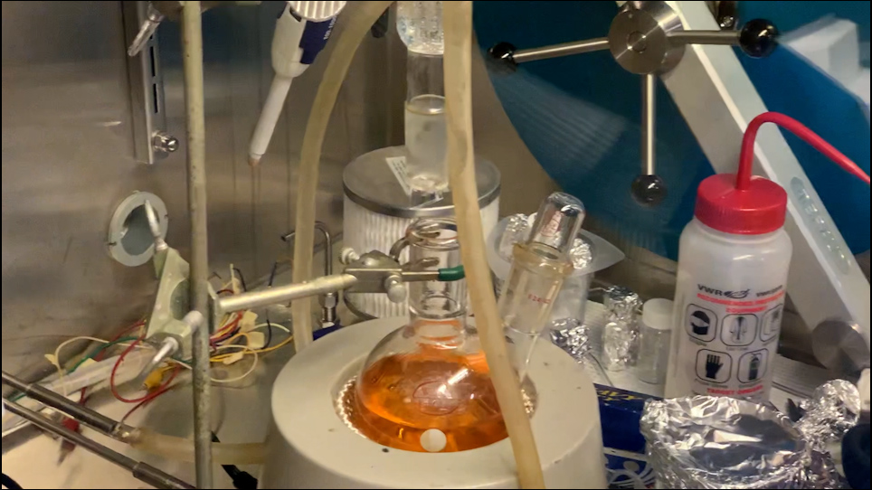
В этом протоколе модифицированные наночастицы из золота, загруженные доксорубицином AS1411-g-PEI-g-PEG, синтезируются с помощью трехступчатых реакций амиде. Затем доксорубицин загружается и доставляется в целевые раковые клетки для лечения рака.

Read Article