JoVE
JoVE
Faculty Resource Center
Research
Behavior
Biochemistry
Biology
Bioengineering
Cancer Research
Chemistry
Developmental Biology
Engineering
Environment
Genetics
Immunology and Infection
Medicine
Neuroscience
JoVE Journal
JoVE Encyclopedia of Experiments
JoVE Chrome Extension
Education
Biology
Chemistry
Clinical
Engineering
Environmental Sciences
Pharmacology
Physics
Psychology
Statistics
JoVE Core
JoVE Science Education
JoVE Lab Manual
JoVE Quiz
JoVE Business
Videos Mapped to your Course
Authors
Librarians
High Schools
About
Sign-In
Sign In
Contact Us
Research
JoVE Journal
JoVE Encyclopedia of Experiments
Education
JoVE Core
JoVE Science Education
JoVE Lab Manual
High Schools
EN
EN - English
CN - 中文
DE - Deutsch
ES - Español
KR - 한국어
IT - Italiano
FR - Français
PT - Português
EN
EN - English
CN - 中文
DE - Deutsch
ES - Español
KR - 한국어
IT - Italiano
FR - Français
PT - Português
Close
Research
Behavior
Biochemistry
Bioengineering
Biology
Cancer Research
Chemistry
Developmental Biology
Engineering
Environment
Genetics
Immunology and Infection
Medicine
Neuroscience
Products
JoVE Journal
JoVE Encyclopedia of Experiments
Education
Biology
Chemistry
Clinical
Engineering
Environmental Sciences
Pharmacology
Physics
Psychology
Statistics
Products
JoVE Core
JoVE Science Education
JoVE Lab Manual
JoVE Quiz
JoVE Business
Videos Mapped to Your Course
Teacher Resources
Get in Touch
Instant Trial
Log In
EN
EN - English
CN - 中文
DE - Deutsch
ES - Español
KR - 한국어
IT - Italiano
FR - Français
PT - Português
Journal
/
Bioengineering
/
用于表征用于超声触发药物释放的荧光标记微气泡的多时间尺度显微镜方法
JoVE Journal
Bioengineering
This content is Free Access.
JoVE Journal
Bioengineering
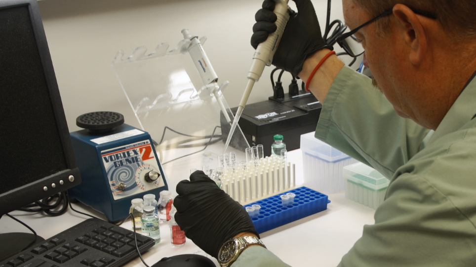
Multi-timescale Microscopy Methods for the Characterization of Fluorescently-labeled Microbubbles for Ultrasound-Triggered Drug Release
Please note that all translations are automatically generated.
Click here for the English version.
用于表征用于超声触发药物释放的荧光标记微气泡的多时间尺度显微镜方法
DOI:
10.3791/62251-v
•
06:02 min
•
June 12, 2021
•
Charlotte Nawijn
,
Tim Segers
2
,
Guillaume Lajoinie
,
Ýrr Mørch
,
Sigrid Berg
5,6
,
Sofie Snipstad
6,7
,
Catharina de Lange Davies
,
Michel Versluis
1
Physics of Fluids group, Department of Science and Technology, MESA+ Institute for Nanotechnology and Technical Medical (TechMed) Center
,
University of Twente
,
2
BIOS Lab-on-a-Chip group, Max Planck Center Twente for Complex Fluid Dynamics, MESA+ Institute for Nanotechnology and Technical Medical (TechMed) Center
,
University of Twente
,
3
Department of Biotechnology and Nanomedicine
,
SINTEF Industry
,
4
Department of Circulation and Medical Imaging
,
Norwegian University of Science and Technology
,
5
Department of Health Research
,
SINTEF Digital
,
6
Cancer Clinic
,
St. Olav’s Hospital
,
7
Department of Physics
,
Norwegian University of Science and Technology
Chapters
00:05
Introduction
00:45
Imaging by Brightfield Microscopy
02:05
Imaging by Fluorescence Microscopy
02:52
Imaging Protocol by Intravital Microscopy
04:07
Results: In Vitro and In Vivo Behavior of Insonified Microbubble
05:22
Conclusion
Summary
Automatic Translation
English (Original)
العربية (Arabic)
中文 (Chinese)
Nederlands (Dutch)
français (French)
Deutsch (German)
עברית (Hebrew)
italiano (Italian)
日本語 (Japanese)
한국어 (Korean)
português (Portuguese)
русский (Russian)
español (Spanish)
Türkçe (Turkish)
Automatic Translation
所提出的方案可用于表征为超声触发的药物递送应用而设计的荧光标记微气泡的反应,包括其激活机制及其生物效应。本文涵盖了一系列
用于
捕获相关长度和时间尺度的
体外和体内
显微镜技术。
Tags
Microbubbles
Ultrasound
Drug Delivery
Fluorescence Microscopy
Brightfield Microscopy
Multi-timescale
Characterization
Article
Embed
ADD TO PLAYLIST
Usage Statistics
Related Videos
单层的合成,装配和表征蛋白质单分子膜的电化学保护的金纳米粒子薄膜
创建的胶粘剂和可溶性梯度荧光显微镜成像细胞迁移
模型和方法来评价药物输送系统在整个细胞屏障的运输
方法表征的共同发展生物膜和生境异质性
交变磁场敏感的混合明胶微凝胶药物控释
用 Widefield 高含量分析系统实现荧光标记组织的自动滑动扫描和分割
用三种互补方法综合评价胎盘靶向药物的有效性和安全性
高通量自动 Microbioreactor 系统在 CHO 细胞模型 IgG1 生产中的应用
用利穆鲁布莱巴细胞裂解液 (lal) 法检测纳米配方中的内毒素
枯草芽孢杆菌
中细菌体和内膜的可视化
Read Article