Journal
/
/
Hydrodynamische Renal bekken injectie voor niet-virale expressie van eiwitten in de nier
JoVE Journal
Biology
A subscription to JoVE is required to view this content.  Sign in or start your free trial.
JoVE Journal Biology
Hydrodynamic Renal Pelvis Injection for Non-viral Expression of Proteins in the Kidney

Hydrodynamische Renal bekken injectie voor niet-virale expressie van eiwitten in de nier

18,060 Views

08:26 min

January 08, 2018

DOI:

08:26 min
January 08, 2018

8 Views
, , , , ,

Transcript

Automatically generated

The overall goal of this surgical procedure is to produce protein expression from plasmid DNA in the adult mouse kidney. This method is a new tool to address key questions in nephrology, such as, how the expression of a protein of interest affects the adult kidney. The main advantage of this technique is that it produces protein expression in a reproducible way, specifically in the kidney, unlike other methods that may also produce gene expression in other organs.

Visual demonstration of this method is critical as injection of the renal pelvis requires careful exposure and must be done quickly in order for the plasma DNA to enter the cells. Start by resuspending plasmid DNA in the hydrodynamic delivery buffer. On a water circulated heat pad covered with a blue pad, place the mouse.

Using a shaver designed for pet grooming, shave the left side of the back of the mouse from shoulder to rump and flank to spine. Clean loose hair and debris and apply vet ointment in both it’s eyes to prevent corneal desiccation. Now, prepare 100 microliters containing 10 to 50 micrograms diluted plasmid DNA in a separate syringe for each mouse that is anesthetized, ensuring that there are no air bubbles.

The needle should be permanently attached without a safety. Prior to performing the surgery, empty sterile surgical tools without touching onto a sterile tray placed on the heat pad. Now, pick up the mouse and lace it in an illuminated field of view.

From the packing, remove 3.15%chlorhexidine gluconate and 70%isopropyl alcohol skin antiseptic swabs and place near the animal. Wear sterile surgical gloves and starting from the site of incision, swab the animal with a new chlorhexidine alcohol swab, three times in a circular motion. Now, locate the incision site.

Pinch the skin with tweezers and using scissors, cut the skin layer approximately one centimeter from the spine, below the ribcage. Make another one centimeter cut below in the muscle layer. Locate the kidney and without touching it, gently expose it from the abdominal cavity by applying a steady gently pressure on the abdomen with the fingers.

Exposing the kidney is sometimes difficult. Apply gentle pressure to the abdomen and spine to avoid damaging the spleen. If organs other than the kidney come out, gently return them to the body cavity using closed forceps if necessary.

Then, gently separate the kidney from the surrounding fat just enough to visualize the renal pelvis, which can be seen as a small white dot. After visualizing the renal pelvis, use closed forceps to push down on the right side of the kidney so that the renal pelvis remains in view. Then, grasp the loaded insulin syringe in the right hand by holding it parallel to the working surface and insert the needle carefully into the renal pelvis.

Once the needle is inserted, inject the solution quickly, within three seconds. Let the needle remain in place for approximately 10 seconds to prevent backflow of the solution. Then, remove the needle slowly and carefully and return the organ to the abdominal cavity by stretching the incision site gently using closed forceps.

Now, suture the muscle layer with five-o absorbable sutures and place two to four independent knots. Similarly, suture the skin layer with five-O or six-O non-absorbable nylon sutures and place two to four independent knots. To assess transfection efficiency, introduce 10 microgram of a luciferase expressing plasmid to the diluted DNA for injection to perform live animal imaging, the following day.

Place the first cage of mice into the chamber and seal it shut. Once anesthetized, inject each mouse intra-peritoneally with 100 microliters of thawed luciferin and record the time. Select the correct user ID and from the top menu bar, click on acquisition and select auto save two from the drop down menu.

Initialize the system by clicking on initialize. Wait for the initializing to finish and change the field of view to D, to image five mice. Ensure that the animals are placed in the desired order and that they’re backs are facing the camera so as to visualize the kidney.

Then, press acquire five minutes following injection to take the image. After acquisition, remove the mice from the imaging machine. Turn off the gas.

Clean the chamber, nose combs and the imaging stage. To analyze the data, after clicking on ROI tools, click the circle icon and select one on the drop down menu to place new ROI in the image as indicated by a red oval with four squares around it. To adjust the size of the ROI, place the cursor over a red square and drag to enclose the purple area overlaying the injected kidney.

Now, right click this ROI and select duplicate ROI to duplicate the ROI in the current window and move it over to the next mouse. After placing all ROI’s from the menu directly above the image, change the units from counts to radiance photons. Finally, create a spreadsheet of the measurements by exporting the ROI data from the tool palate by clicking on ROI tools and then on the pencil ruler icon.

The kidney’s specific gene expression of reporter plasmids is shown. All mice were injected with pTet plasmid expressing enhanced firefly luciferase, while mice two, three and five were also injected with pEF1-alpha-tdtomato plasmid, expressing the red fluorescent protein, tdtomato. Generally, the radiance in young mice will be within one order of magnitude.

It is possible that some mice do not show appreciable levels of radiance, either due to low transfection efficiency or lack of the transgene itself. Injection of hydrodynamic buffer serves as a control. No plasmid expression is observed when given only the hydrodynamic buffer.

In a first few days following transfection, there will likely be a low number of cells staining positive for the transgene. These cells are seen localized in patches in the areas infiltrated by the injection fluid. In this image, the green signals are from the proximal tubules, which are stained for lotus tetragonolobus agglutinin and the red signals are from the transfected cells expressing the tdtomato reporter gene.

Once mastered, the surgery can be done in less than 10 minutes, however, the anesthesia takes time to induce and recover from. So, two trained personnel, working closely together would be able to do only about 10 to 15 mice per day. While attempting this procedure, it’s important to remember to use young mice.

The hydrodynamic technique does not work as well in older fibrotic organs. Following this procedure, other methods can be performed, such as analysis of blood, urine and kidney tissue samples, in order to assess the expression of the gene of interest. After watching this video, you should have a good understanding of how to inject the renal pelvis with plasmid DNA to achieve kidney specific protein expression.

Summary

Automatically generated

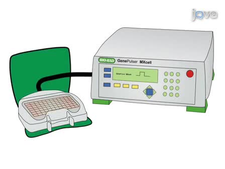
Dit protocol beschrijft een methode om te injecteren plasmide DNA in de nier van de muis via de renal bekken te produceren transgenic expressie specifiek in de nier.

Read Article