In vitro spheres assays are commonly used to identify cancer stem cells. Here we compare single with multi cell-based spheres assays. The more laborious single cell-based assays or methylcellulose supplementation give more accurate results while multi cell-based assays performed in liquid medium can be highly influenced by cell density.
Years of research indicates that ovarian cancers harbor a heterogeneous mixture of cells including a subpopulation of so-called “cancer stem cells” (CSCs) responsible for tumor initiation, maintenance and relapse following conventional chemotherapies. Identification of ovarian CSCs is therefore an important goal. A commonly used method to assess CSC potential in vitro is the spheres assay in which cells are plated under non-adherent culture conditions in serum-free medium supplemented with growth factors and sphere formation is scored after a few days. Here, we review currently available protocols for human ovarian cancer spheres assays and perform a side-by-side analysis between commonly used multi cell-based assays and a more accurate system based on single cell plating. Our results indicate that both multi cell-based as well as single cell-based spheres assays can be used to investigate sphere formation in vitro. The more laborious and expensive single cell-based assays are more suitable for functional assessment of individual cells and lead to overall more accurate results while multi cell-based assays can be strongly influenced by the density of plated cells and require titration experiments upfront. Methylcellulose supplementation to multi cell-based assays can be effectively used to reduce mechanical artifacts.
There is increasing evidence that ovarian carcinomas are comprised of heterogeneous mixtures of cells and harbor so-called “cancer stem cells” (CSCs) responsible for disease initiation, maintenance and relapse after conventional cytotoxic therapies1-3. Therefore, the development of molecular strategies targeting ovarian CSCs is an important goal and promises to improve the therapy of ovarian cancer patients.
A pre-requisite for the understanding of the molecular features of CSCs is their reliable isolation from the non-CSCs. However, identification of ovarian CSCs appears challenging. While CD133 expression and aldehyde dehydrogenase (ALDH) activity4,5 have been reported to mark ovarian CSCs, some data indicate that these markers are unstable6. Consistently, in ovarian cancer, other than for example in breast carcinoma7, expression of ALDH1 associates with favorable outcome8 and expression of the proposed stem cell marker CD44 variant has no prognostic value9. More recently, we have shown that expression of the embryonic stem cell protein SOX2 confers stemness to ovarian carcinoma cells10 and high SOX2 expression associates with clinically aggressive ovarian and breast carcinomas11,12. Therefore, in this report we use a lentiviral reporter construct containing a red fluorescence protein (RFP) whose expression is controlled by a SOX2 regulatory region, as a method to isolate putative ovarian CSCs.
By definition, CSCs can both self-renew and differentiate, giving rise to all tumor cell types. Putative CSC populations need to be analyzed in functional assays performed in vivo. For obvious reasons, in human cells such functional tests are confined to xenograft assays, comprising mostly transplantation of human tumor cells into immuno-compromised mice10,13.
An alternative in vitro method was offered by Brent Reynolds and Sam Weiss who firstly reported the so-called neurosphere assay as a surrogate assay evaluating stem potential in neural cells14. Dontu and colleagues later confirmed the use of this assay for evaluation of stem cell potential in breast cells15,16. Here, human mammary cells were plated in different numbers in serum-free medium supplemented with epidermal growth factor (EGF), basic fibroblast growth factor (bFGF), B-27 and heparin and cultured under non-adherent conditions for seven to ten days before sphere formation was scored by microscopy. Following this protocol with some adjustments in cell numbers, growth medium and supplements, several groups have explored in vitro stem cell potential from several cancer types such as breast17, brain18, pancreas19 and colon20 tumors. In ovarian carcinoma, we have recently reported feasibility of the spheres assay and compared its results to those collected in in vivo murine xenograft models10. We found that overexpression of the stem cell protein SOX2 enhanced both in vitro sphere formation as well as in vivo tumorigenicity of human ovarian carcinoma cells10. However, the frequency of sphere-initiating cells was higher than the frequency of tumor-initiating cells measured in vivo10 suggesting that either the sphere assay may lead to false positive results due to technical reasons or, alternatively, the in vivo assay may be inefficient and result in false negative results.
In this report, we analyze multi cell-based ovarian spheres assays in more detail, review the different protocols available in the literature and compare them to a single cell-based assay. We show that the single cell-based assay provides more accurate and reproducible results than multi cell-based assays, which can be highly influenced by the density of plated cells unless methylcellulose is added to the cultures to immobilize cells. However, also in single cell-based assays, in vitro sphere-initiating potential is observed at higher frequency than in vivo tumor-initiating potential.
1. Generation of OVCAR-3 Human ovarian Carcinoma Cells Stably Transduced with Lentiviruses Containing the SOX2 Regulatory Region Reporter Construct
2. Preparation of Cell Sorting and Plating
3. Serial Passaging of Spheres
4. Result Analysis
In conventional spheres assays, nearly 40% of RFP+ OVCAR-3 cells vs. 20% of RFP- cells gave rise to an individual tumor sphere in the primary spheres assay (Figure 4A). Moreover, spheres formed by RFP+ cells were larger in size than those formed by RFP- cells.
When plated in single cell-based assays, RFP+ cells also formed more spheres than RFP- cells, confirming the results above. However, there was a tendency towards generation of fewer spheres per plated in the single versus the multi cell-based assay (Figure 4A,B), indicating that in this assay sphere formation may be biased through technical artifacts such as mechanical sphere fusion or dissociation in the non-adherent culture medium.
To further explore these aspects, we compared the influence of cell plating density on spheres formation. We plated cells using limiting dilution from 1,000 cells to 1 cell per well in 96-well plates and found that the numbers of emerging spheres were highly dependent on the numbers of initially plated cells. Surprisingly, higher numbers of spheres were counted from lower numbers of plated cells in both MGEM and DMEM/F12-based media, demonstrating that indeed plating modalities highly bias results in this assay (Figure 5). In contrast, when cells were immobilized by adding 1% methylcellulose to DMEM/F12-based spheres medium23,24 the efficiency of sphere formation was mostly independent of cell density.
To explore the influence of spheres culture conditions on CSC properties, we analyzed the percentage of cells expressing red fluorescence signal after 7 days of incubation in the spheres assay. We found that in multi cell-based spheres cultures approximately 35% of the cells from RFP+ spheres lost their red fluorescence signal after seven days of culture (Figure 6A), suggesting that they have undergone differentiation, while 65% retained a RFP+ signal suggesting self-renewal capacity. In contrast, cells from spheres generated from initially RFP- cells remained RFP negative (Figure 6A), indicating that they cannot re-establish stem cell potential under these conditions.
Fluorescence microscopy performed on spheres generated from single cells confirmed these results showing that single spheres derived RFP+ cells contained both RFP+ and RFP- cells while spheres derived from RFP- cells remained negative for the red signal.
Similar results were observed in replating assays from both conditions.
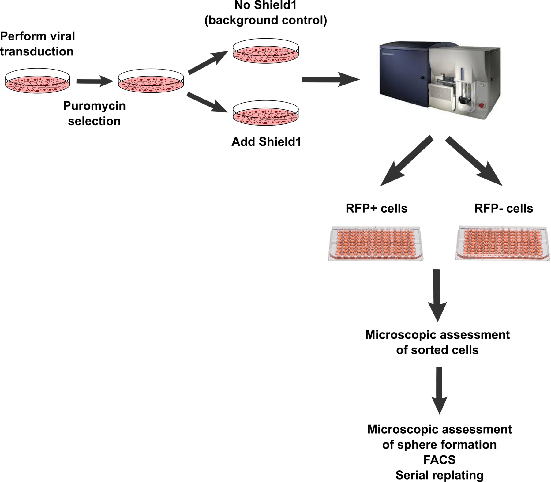
Figure 1. Workflow of lentiviral transduction, selection and sorting of RFP+ and RFP- cells. After lentiviral transduction and positive selection of successfully transduced cells via puromycin exposure, RFP- and RPF+ cells are sorted by FACS into individual wells of a 96-well plate in spheres medium. For multi cell-based spheres assays, 100 cells are placed into one well. Plating efficiency is assessed by microscopy performed after sorting. Spheres were scored by microscopy after seven to ten days, dissociated into single cells, analyzed via flow cytometry and replated into secondary spheres. Please click here to view a larger version of this figure.
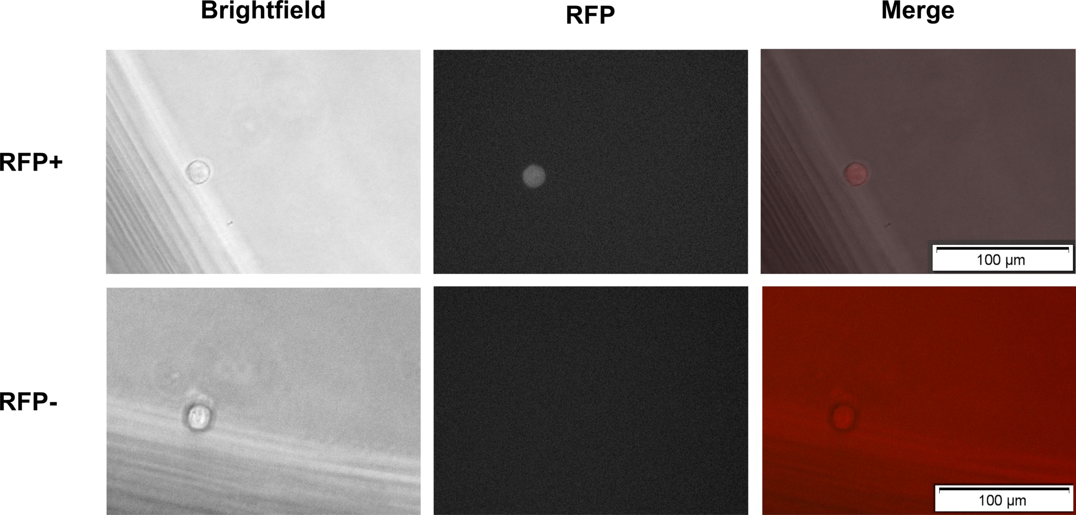
Figure 2. Imaging of sorted RFP+ and RFP- cells after plating. Single RFP+ and RFP- cells sorted into each well of a 96-well plate are analyzed for correct plating by using a (fluorescent) microscope. Please click here to view a larger version of this figure.
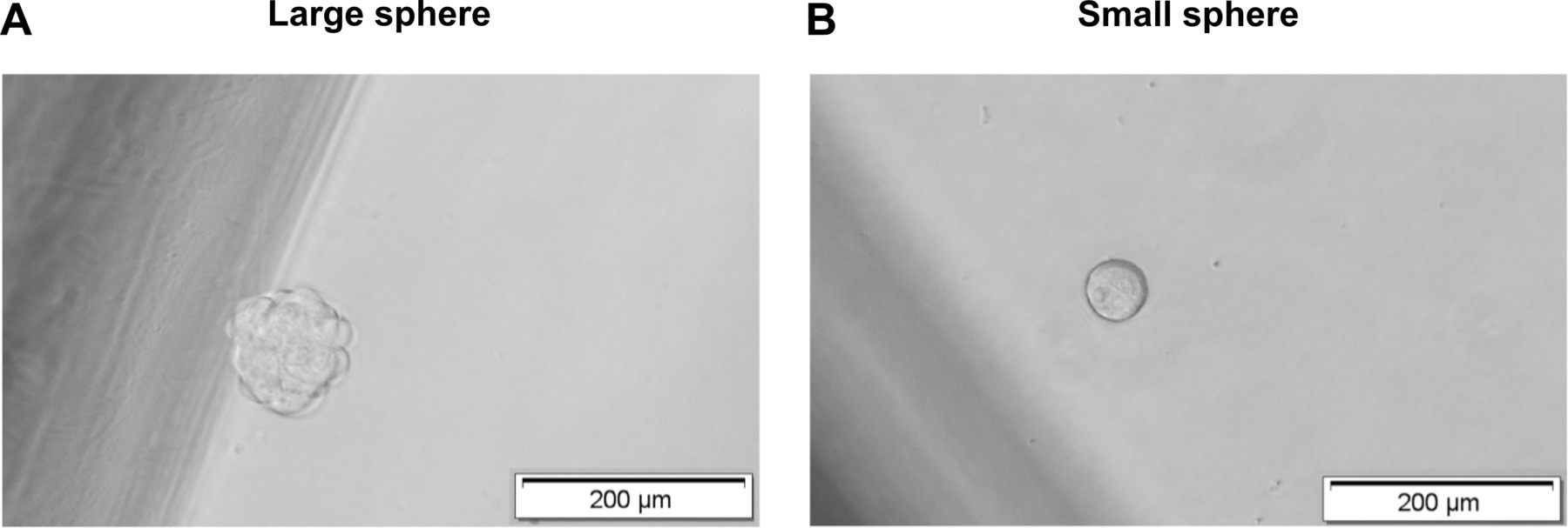
Figure 3. Analysis of spheres formation in single cell-based assays. Spheres formation is analyzed after seven to ten days. (A) Large (diameter > 100 µm) and (B) small (diameter 50 – 100 µm) spheres are distinguished microscopically based on size. Please click here to view a larger version of this figure.
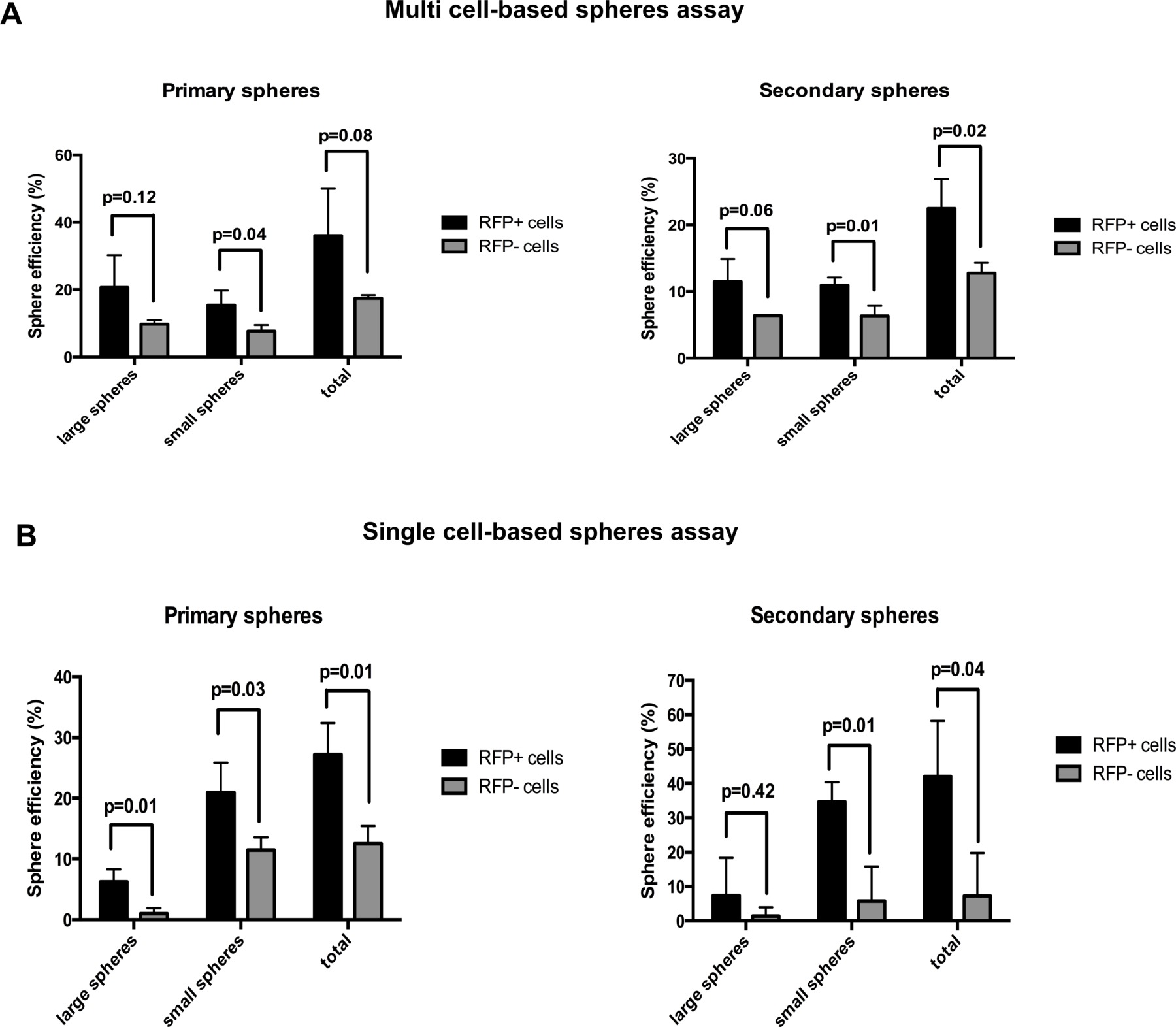
Figure 4. Efficiency of tumor spheres formation from RFP+ and RFP- OVCAR-3 cells in multi versus single-cell based spheres assays. Comparison of primary and secondary sphere efficiency from OVCAR-3 cells as assayed in multi (A) versus single cell-based spheres assays (B). Please click here to view a larger version of this figure.
Figure 5. Cell plating density strongly impacts sphere counts from OVCAR-3 cells in the multi cell-based spheres assay performed in liquid but not in methylcellulose supplemented cultures. Use of different cell densities and growth media have been reported in the literature for ovarian cancer spheres assays. To analyze possible biases introduced by these variables, cells are plated at different densities in 200 µl of different spheres culture media (MGEM, DMEM/F12 with all supplements as detailed in the protocol section, or DMEM/F12 with all supplements and containing 1% methylcellulose) and sphere formation is scored after 7 days (A). Shown in (B) are microscopy pictures of cells plated at different densities taken one day after plating in DMEM/F12 spheres culture medium without methylcellulose. Note the cell clusters emerging at high cellular density as opposed to single cells seen in low density plates. Scale bar for pictures: 50 µm. Please click here to view a larger version of this figure.
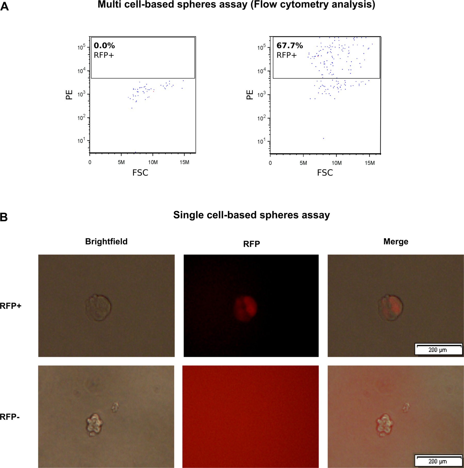
Figure 6. Analysis of RFP signal in tumor spheres formed from RFP+ and respectively RFP- OVCAR-3 cells. (A) Flow cytometry analysis for RFP signal in dissociated spheres derived from RFP+ and RFP- cells (multi cell-based spheres assay); (B) Microscopy of spheres derived from RFP+ and RFP- cells (single cell-based spheres assay) reveals heterogeneous RFP signal in spheres derived from RFP+ but not from RFP- cells. Pictures were taken at day 7 for conventional spheres and day 10 for single cell-based sphere assays. Note the larger size of spheres derived from RFP+ putative CSCs. Please click here to view a larger version of this figure.
Human ovarian cancer cell source | Basic medium | Supplements | Authors |
OVCAR-3, Caov-3, primary material | MEGM | 20 ng/ml rEGF, 20 ng/ml bFGF, B-27, 4 μg/ml heparin, hydrocortisone, insulin (SingleQuot kit) | Bareiss et al. |
SKOV3 | DMEM/F12 | 5 µg/ml insulin, 10 ng/ml rEGF, 10 ng/ml bFGF, 12 ng/ml LIF, 0.3% BSA | Li Ma et al. |
A2780 | DMEM/F12 | 5 µg/ml insulin, 20 ng/ml rEGF, 2% B-27, 0.4% BSA | Haiwei Wang et al. |
SKOV3 | DMEM/F12 | 5 µg/ml insulin, 20 ng/ml rEGF, 10 ng/ml bFGF, 2% B-27, 1 ng/ml hydrocortisone | Yong-Rui Du et al. |
A2780, primary material | DMEM/F12 | 5 µg/ml insulin, 20 ng/ml rEGF, 10 ng/ml bFGF, 0.4% BSA | T. Xiang et al. |
Primary material | DMEM/F12 | 5 µg/ml insulin, 10 ng/ml rEGF, 10 ng/ml bFGF, 12 ng/ml LIF, 0.3% BSA | Te Liu et al. |
MLS | DMEM/F12 | 10 ng/ml insulin, 20 ng/ml rEGF, 20 ng/ml bFGF, 2% B-27 | Soritau et al. |
3AO | DMEM/F12 | 1 mg/ml insulin, 20 ng/ml rEGF, 20 ng/ml bFGF, 2% B-27 | M. F. Shi et al. |
Primary material | DMEM/F12 | 5 µg/ml insulin, 20 ng/ml rEGF, 10 ng/ml bFGF, 0.4% BSA | Shu Zhang et al. |
Primary material | EBM-2 or X-VIVO | 5 µg/ml insulin, 20 ng/ml rEGF | Ilona Kryczek et al. |
OVCAR-3 | MEGM | 20 ng/ml rEGF, 20 ng/ml bFGF, B-27, 4 μg/mL heparin | Dongming Liang et al. |
Table 1. Examples of different cell sources (cell lines and primary patient-derived tissue), media and supplements used for ovarian spheres assays in previous reports.
Spheres cultures are a widely used method to assay cancer stem cell potential and enrich for stem-like cells in a wide range of human tumor cells15,25,26. Under these culture conditions, cancer cells that lack self-renewal ability are expected to differentiate and eventually undergo cell death. Although they may initially form cell clusters or even tumor spheres especially in primary assays, they are not able to sustain sphere-forming ability upon serial replating due to lack of self-renewing properties. Spheres assays are used as surrogate assays to identify CSCs and evaluate their frequency in whole tumor cell populations.
However, substantial variability can be observed between spheres assays performed following different published protocols5,10,27-35 (Table 1). In our laboratory, we have previously published sphere formation from human ovarian carcinoma cells using MEGM supplemented with B-27, bFGF, Heparin and SingleQuotTM (containing insulin, rEGF and hydrocortisone). Other labs use whole DMEM/F12 Medium, while some add only B-27 and rEGF. In this report, multi cell-based spheres assay in OVCAR-3 cells were therefore performed using different conditions. Using MEGM or DMEM/F12 with all supplements no significant difference in sphere formation was observed in these cells (Figure 5A). In addition, some labs have speculated that EGF and FGF may be quickly degraded and have established protocols adding these growth factors daily to the medium. We therefore compared spheres assays performed in a medium in which EGF and FGF was added only at the beginning of the spheres assays with daily addition of EGF and FGF to the cell culture, and we find these assays to yield equivalent results in OVCAR-3 cells (data not shown), suggesting that the expensive and laborious daily supplementation with EGF and FGF may not always be necessary. Whether these results are applicable to cells from other ovarian cancer cell lines or primary samples, or under different experimental conditions remains to be determined.
However, we observed a substantial bias in the numbers of scored spheres introduced by another tested variable, the cell plating density. Surprisingly, wells seeded with lower numbers of cells showed higher numbers of spheres. Limiting cell mobility by 1% methylcellulose resulted in the same efficiency of sphere formation, independent on the number of initially plated cells. These results suggest that cell clumping and sphere fusion or disaggregation can occur modifying sphere numbers and leading to inaccurate results in multi cell-based spheres assays. When cells are plated at proper density, multi cell-based assays however lead to results rather comparable to data collected in single cell-based sphere assays (Figure 4). To further explore these results we compared single and multi cell-based assays using ovarian carcinoma cells sorted into putative CSCs via a recently published lentiviral RFP expressing reporter system for a SOX2 regulatory region10. Indeed, both assays confirmed the enhanced primary and secondary sphere forming capacity of RFP+ versus RFP- cells (Figure 4A,B). Importantly, the higher numbers of spheres observed from RFP+ cells were not due to higher proliferative capacity of the SOX2 expressing cells (data not shown), which is in line with previous results showing that induction of SOX2 promotes spheres formation and in vivo tumorigenicity without accelerating cell cycle progression10.
Taken together, single cell-based spheres assays are more laborious and expensive but they result in more accurate data, which also is confirmed by the higher reproducibility of results between experiments. Since plating density highly influences results in multi cell-based suspension spheres assays, upfront titration of adequate plating density is required for each individual tumor cell type before assaying sphere formation using these assays. Alternatively, the more accurate single cell-based assays can be used upfront, or methylcellulose supplementation to improve accuracy of results by reducing mechanical artifacts. If sphere formation is compared between conditions where the genetic modification or drug treatment may severely alter viability of the cells, thereby decreasing cell density, single cell-based sphere assays may be mandatory to avoid false positive results.
In summary, under proper experimental conditions both the multi cell-based spheres assay and the single cell-based spheres assay are able to indicate differences in sphere potential between different cell populations (stem and non-stem cells). However, multi cell-based spheres assays which commonly are performed in liquid cultures are more susceptible to errors introduced by experimental design through plating density. Supplementation of methylcellulose (1%) to multi cell-based assays can limit artifacts related to cell clumping and sphere fusion. Based on these data, we recommend single cell-based spheres assays to be performed unless detailed titration analyses have been performed upfront and negative impact of experimental conditions on cell viability and thereby cell density has been ruled out. However, single cell-based spheres assays are more laborious and more expensive, and might not be required in each experimental setting. Methylcellulose-supplemented multi cell-based spheres assays may represent another alternative in some experimental settings.
The authors have nothing to disclose.
This study was supported by a grant from the Baden-Württemberg Stiftung (Adult Stem Cells Program II) awarded to C.L. We thank Dr. Martina Konantz for critical input and review of the manuscript. We thank Emmanuel Traunecker and Toni Krebs from the DBM FACS Facility (University Hospital Basel) for assistance with FACS sorting.
Name of Material/ Equipment | Company | Catalog Number | Comments/Description |
low-Attachment-plate | Corning | 3474 | |
MEGM | Lonza | CC-3151 | |
Insulin | Lonza | CC-4136 | SingleQuots™ Kit |
Hydrocortison | Lonza | CC-4136 | SingleQuots™ Kit |
EGF | Lonza | CC-4136 | SingleQuots™ Kit |
EGF | Sigma | E9644 | end concentration: 20 ng/ml |
FGF | PeproTech | 100-18B | end concentration: 20 ng/ml |
B-27 | Invitrogen/ Gibco | 17504-044 | end concentration: 1x |
Heparin-Natrium-25000 IE | Ratiopharm | N68542.02 | dilution 1:1000 |
Pen/Strep | Gibco | 15140-122 | |
FCS | Gibco | 10500-064 | |
RPMI 1640 | Gibco | 21875-034 | |
Trypsin-EDTA | Gibco | 25300-054 | |
Dulbecco’s PBS (1x) | Gibco | 14190-094 | |
Shield1 | Clontech | 632189 | dilution 1:1000 |
DMEM/F12 | Gibco | 21041-025 | |
DMEM/F12 (powder) | Gibco | 42400-010 | |
Methyl cellulose | Sigma | M0387 | |
Puromycin dihydrochloride | applichem | A2856 | |
cell sorter | BD | Aria III cell sorter | |
FACS analyser | BD | accuri c6 flow cytometer | |
microscope | Olympus | IX50 Osiris |