For CHS induction, the animals were sensitized via skin painting (abdominal) with 150 µL of 5% TNCB or sham sensitized with the vehicle alone. On day "4", the ear swelling responses of both ears were induced by contact painting (challenge) with 10 µL of 0.4% TNCB in both mice previously contact sensitized with TNCB (test group) and the control group mice (sham sensitized). The presented data depict that the mice sensitized with TNCB and challenged 4 days later developed significantly increased ear swelling compared with the sham-sensitized ones similarly challenged (Figure 3, Table 2, test vs. control group). The ear swelling results were completely validated in further studies, highlighting that an increase in ear edema determined with a micrometer agreed with augmented ear weight (Figure 4), MPO activity (Figure 5), IFN-γ concentration in the ear extracts (Figure 6), increased thickening of the edematous dermis in the histological examination (Figure 7), and ear vascular permeability (Figure 8). An increase in the concentration of TNP-specific IgG1 antibodies was also found in the sera of the test mice when compared with the control animals (Figure 9).
As an example of T cell-mediated immune response, CHS can also be transferred into naïve syngeneic recipient mice. Donors were sensitized by TNCB application, and subsequently, the CHS-effector cells were administered i.v. into the naïve recipient mice, which were challenged with the hapten and tested for CHS 24 h later (Figure 10). The animals that received the CHS-effector cells from donors previously sensitized with TNCB showed significantly increased ear swelling compared to animals that were challenged only (did not receive any cells).
The CHS reaction has a complex mechanism and involves various cells. Antigen presentation and T/B cell activation occur in the peripheral lymph organs (e.g., ALNs and SPL). It was determined that CHS-effector cells depleted of CD4+ but not CD8+ cells prior to adoptive cell transfer resulted in the absence of the CHS reaction in the recipient mice. Those cells were found to be positive for IFN-γ (T-box transcription factor TBX21, Tbet+) and IL-17A (retinoic acid receptor-related orphan nuclear receptor gamma, RORγT+) (Supplementary Figure 1).
The presented results from the representative experiments were performed on C57BL/6 and CBA/J male and female mice at 8-12 weeks of age. Following the 3R rules in the use of animals23, especially reduction, for the purposes of this article, the results of experiments are shown on small groups of animals. Data in the graphs are shown as mean ± SEM. Statistical significance was set at p < 0.05. The graphs were drawn using Prism software (see Table of Materials).
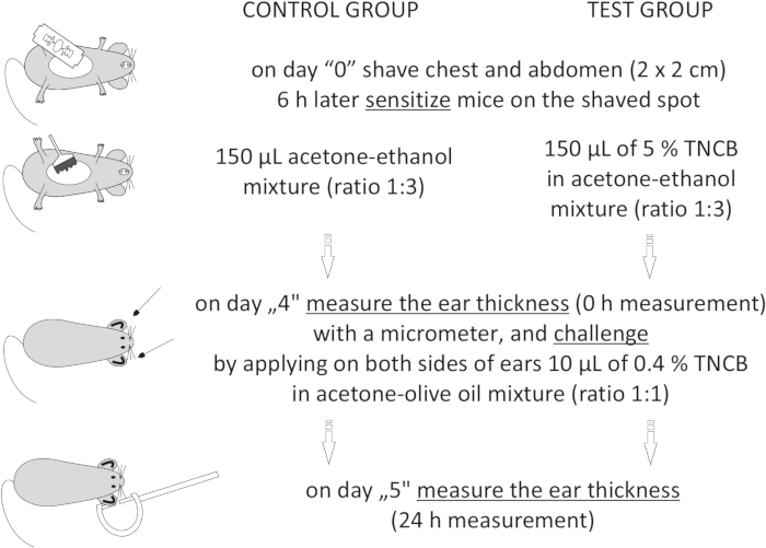
Figure 1: Induction of CHS. Sensitization, challenge, and ear measurement. Abbreviations: CHS = contact hypersensitivity reaction; TNCB = 2,4,6-trinitrochlorobenzene. Please click here to view a larger version of this figure.
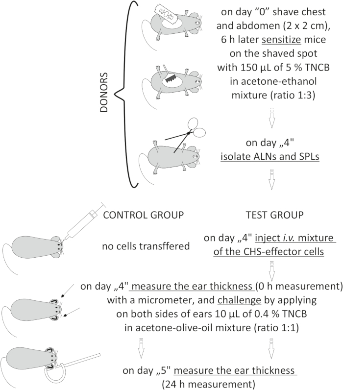
Figure 2: Adoptive transfer of the CHS-effector cells. Abbreviations: ALNs =axillary and inguinal lymph nodes; CHS = contact hypersensitivity reaction; i.v. = intravenously; SPLs = spleens; TNCB = 2,4,6-trinitrochlorobenzene. Please click here to view a larger version of this figure.

Figure 3: Representative evaluation of CHS to TNCB by the measurement of ear swelling with a micrometer. Mice were TNCB (test group) or sham (control group) sensitized and subsequently challenged. The thickness of the auricle was measured before and after the challenge, and differences in ear swelling were calculated by subtracting the 0 h ear thickness (µm) from the 24 h ear thickness (µm). Ear swelling was expressed as mean ± SEM, ****p < 0.0001, n = 10 mice/group (data from Table 2). Abbreviations: CHS = contact hypersensitivity reaction; SEM = standard error of the mean; TNCB = 2,4,6-trinitrochlorobenzene. Please click here to view a larger version of this figure.

Figure 4: Representative evaluation of CHS by the measurement of ear weight. Ear weight is one of the parameters that corresponds with ear swelling. Mice were TNCB (test group) or sham (control group) sensitized and subsequently challenged. At 24 h after the challenge, 6 mm diameter punches were taken from the removed ears. The punches were weighed on an analytical laboratory balance. Ear weight was expressed in milligrams (mg) as mean ± SEM, ***p < 0.001, n = 10 mice/group. Abbreviations: CHS = contact hypersensitivity reaction; SEM = standard error of the mean; TNCB = 2,4,6-trinitrochlorobenzene. Please click here to view a larger version of this figure.

Figure 5: Representative evaluation of MPO activity. Increased MPO activity in tissue extracts correlates with ear inflammation. TNCB-sensitized mice (test group) and sham-sensitized mice (control group) were challenged. At 24 h post-challenge, the ears were removed, and 6 mm diameter punches of the ear were extracted and processed. MPO activity is expressed in U per protein content (U/g of protein). Results displayed as mean ± SEM, **p < 0.01, n = 5-6 mice/ group. Abbreviations: CHS = contact hypersensitivity reaction; MPO = myeloperoxidase; SEM = standard error of the mean; TNCB = 2,4,6-trinitrochlorobenzene; U = units. Please click here to view a larger version of this figure.

Figure 6: Representative evaluation of cytokine production-IFN-γ concentration in ear extracts. Mice were TNCB (test group) or sham (control group) sensitized and subsequently challenged. At 24 h post-challenge, the ears were removed, and 6 mm diameter punches of the ear were taken. The concentration of IFN-γ was determined in tissue homogenates by ELISA. Results shown as mean ± SEM, *p < 0.05, n = 5 mice/group. Abbreviations: CHS = contact hypersensitivity reaction; IFN-γ = interferon gamma; SEM = standard error of the mean; TNCB = 2,4,6-trinitrochlorobenzene. Please click here to view a larger version of this figure.

Figure 7: Representative histology of the ear tissue. Hematoxylin and eosin staining. Mice were TNCB (test group) or sham (control group) sensitized and subsequently challenged. (A–C) Histological examination in the test group manifested in a significantly increased concentration of inflammatory cells (mononuclear and polymorphonuclear cells), mainly in the dermis, with microabscess formation in the epidermis. Thickening of the edematous dermis and a thickened, hyperplastic epidermis were also noticed. (D–E) Control group. Please click here to view a larger version of this figure.

Figure 8: Representative vascular permeability test. Observed ear tissue edema is a result of increased vascular permeability. To determine changes in vascular permeability, mice were TNCB (test group) or sham (control group) sensitized and then challenged 4 days later. At 23 h after the challenge, Evans blue was injected, and, 1 h after Evans blue injection, the animals were euthanized, and 6 mm diameter punches of the ear were made. Results shown as mean ± SEM, **p < 0.01, n = 5 mice/group. Abbreviations: CHS = contact hypersensitivity reaction; SEM = standard error of the mean; TNCB = 2,4,6-trinitrochlorobenzene. Please click here to view a larger version of this figure.

Figure 9: Representative anti-TNP IgG1 antibody measurement. The concentration of anti-TNP IgG1 antibodies in serum was measured 24 h after the challenge with hapten TNCB in sham sensitized (control) and in TNCB-sensitized (test group) mice. The collected sera were tested for antibody concentration by ELISA. Results shown as mean ± SEM, ***p < 0.001, n = 10 mice/group. Abbreviations: CHS = contact hypersensitivity reaction; IgG1 = immunoglobulin G subclass 1; SEM = standard error of the mean; TNCB = 2,4,6-trinitrochlorobenzene. Please click here to view a larger version of this figure.

Figure 10: Representative adoptive transfer of the CHS-effector cells. The CHS-effector cells were obtained from donors that were sensitized with TNCB. Next, the collected immune cells were injected, i.v., into naïve syngeneic recipients, which were challenged for elicitation of the CHS effector phase. The control group of mice did not receive any cells prior to the challenge. The thickness of the auricle was measured before and after the challenge. Results shown as mean ± SEM, ***p < 0.001, n = 7 mice/group. Abbreviations: CHS = contact hypersensitivity reaction; SEM = standard error of the mean; TNCB = 2,4,6-trinitrochlorobenzene. Please click here to view a larger version of this figure.
| Mouse strain | Sensitization solution (dose) on shaved abdomen |
Elicitation solution (dose) on both sides of ear/ s |
Sensitization / elicitation day | Refs | |||||||||||
| BALB/c (H-2d); C57BL/6 (H-2b) TCRδ-/-, β2m-/-, CD1d-/- (B10 PL (H-2u background) |
25 μL of 0.5 % DNFB in acetone-olive oil mixture (ratio 4:1) | 5 μL of 0.1 % DNFB in acetone-olive oil mixture (ratio 4:1) | 0 / 5 | 22 | |||||||||||
| C57BL/6 (H-2b) | 50 μL of 0.5 % DNFB in acetone-olive oil mixture (ratio 4:1) | 25 μL of 0.2 % DNFB in acetone-olive oil mixture (ratio 4:1) | 0 / 5 | 32 | |||||||||||
| C57BL/6 (H2b); IL-17A-/- (C57BL/6 background) | 150 μL of 5% TNCB in acetone-ethanol mixture (ratio 1:3) | 10 μL of 0.4 % TNCB in olive oil-acetone mixture (ratio 1:1) | 0 / 4 | 33 | |||||||||||
| CBA/J (H-2k); C57BL/6 (H-2b) TLR2-/-, MyD88-/-, IL-17A-/- (C57BL/6 background) |
150 μL of 5 % TNP-Cl (TNCB) in an acetone-ethanol mixture (ratio 1:3) | 10 μl of 0.4 % TNP-Cl (TNCB) in olive oil-acetone mixture (ratio 1:1) | 0 / 4 | 21 | |||||||||||
| C57BL/6 (H-2b); BALB/c (H-2d) | 25 μL of 1 % TNCB in an acetone | 10 μL of 0.1 or 0.2 % TNCB in an acetone (and higher up to 1 %) | 0 / 7 | 34 | |||||||||||
| C57BL/6 (H-2b); TLR2-/-/ TLR4-/- (double-knockout mice on C57BL/6 background) |
100 μL of 3 % TNCB in an acetone | 20 μL of 1 % TNCB in an acetone (just on the back side of ears) | 0 / 5 | 31 | |||||||||||
| C57BL/6 (H-2b) MHC class II-deficient mice (C57BL/6 background) |
100 μL of 3 % TNCB in acetone-olive oil mixture (ratio 4:1) | 20 μL of 0.5 or 1 % TNCB in acetone-olive oil mixture (ratio 4:1) | 0 / 6 | 35 | |||||||||||
| C57BL/6 (H-2b) | 100 μL of 7 % TNCB in an acetone | 20 μL 1 % TNCB in an acetone | 0 / 5 | 36 | |||||||||||
| C57BL/6 (H-2b) | 100 μL 3 % OX in ethanol | 20 μL 1 % OX in an ethanol | 0 / 5 | ||||||||||||
| C57BL/6 (H-2b); CD4-/-, CD8-/- (C57BL/6 background) | 25 μL of 0.5 % DNFB in acetone-olive oil (ratio 4:1) | 10 μL of 0.2 % DNFB in acetone-olive oil (ratio 4:1) | 0 / 5 | 10 | |||||||||||
| C57BL/6 (H-2b); CD4-/-, CD8-/- (C57BL/6 background) | 150 μL of 3 % OX in alcohol-acetone (ratio 3:1) | 10 μL 1% OX in alcohol-acetone (ratio 3:1) | 0 / 5 | ||||||||||||
| C57BL/6 (H-2b); C3H/HeN (H-2k); TLR4-/- (C3H/HeJ background); MyD88-/- (C57BL/6 background) | 100 mg/ ear of 10 % NiCl2 in white petrolatum on the dorsal side of both ears | 10 % NiCl2 in white petrolatum | 0,1,2 / 23, 24 | 37 | |||||||||||
| NOD (H-2g7) MyD88-/- (NOD background) | 400 μL of 0.5 % FITC in acetone and dibutyl phthalate | 10 μL of 0.1 % FITC in acetone and dibutyl phthalate | 0 / 5 | 25 | |||||||||||
Table 1: Comparison of the CHS model in various studies. Abbreviations: DNFB = 1-fluoro-2,4-dinitrobenzene; FITC = fluorescein isothiocyanate; NiCl2 = nickel (II) chloride; TNCB = 2,4,6-trinitrochlorobenzene; TNP-Cl = trinitrophenyl chloride; OX = oxazolone. Please click here to download this Table.
| Control group (negative) | Test group (CHS reaction) | ||||||
| Mouse # ear: L, R | 0 h ear thickness [μm] | 24 h ear thickness [μm] | 24 h – 0 h ear thickness [μm] | Mouse # ear: L, R | 0 h ear thickness [μm] | 24 h ear thickness [μm] | 24 h – 0 h ear thickness [μm] |
| 1 L | 365 | 380 | 15 | 1 L | 345 | 427.5 | 82.5 |
| 1 R | 335 | 380 | 45 | 1 R | 340 | 455 | 115 |
| 2 L | 345 | 355 | 10 | 2 L | 355 | 475 | 120 |
| 2 R | 327.5 | 352.5 | 25 | 2 R | 342.5 | 457.5 | 115 |
| 3 L | 340 | 370 | 30 | 3 L | 340 | 460 | 120 |
| 3 R | 325 | 355 | 30 | 3 R | 345 | 495 | 150 |
| 4 L | 335 | 380 | 45 | 4 L | 357.5 | 432.5 | 75 |
| 4 R | 340 | 350 | 10 | 4 R | 335 | 402.5 | 67.5 |
| 5 L | 350 | 380 | 30 | 5 L | 335 | 387.5 | 52.5 |
| 5 R | 337.5 | 360 | 22.5 | 5 R | 335 | 425 | 90 |
| 6 L | 335 | 365 | 30 | 6 L | 350 | 430 | 80 |
| 6 R | 340 | 375 | 35 | 6 R | 342.5 | 405 | 62.5 |
| 7 L | 345 | 337.5 | 0 | 7 L | 340 | 502.5 | 162.5 |
| 7 R | 345 | 335 | 0 | 7 R | 327.5 | 447.5 | 120 |
| 8 L | 370 | 380 | 10 | 8 L | 327.5 | 515 | 187.5 |
| 8 R | 375 | 355 | 0 | 8 R | 327.5 | 540 | 212.5 |
| 9 L | 385 | 370 | 0 | 9 L | 330 | 415 | 85 |
| 9 R | 342.5 | 362.5 | 20 | 9 R | 327.5 | 390 | 62.5 |
| 10 L | 307.5 | 340 | 32.5 | 10 L | 337.5 | 445 | 107.5 |
| 10 R | 325 | 350 | 25 | 10 R | 352.5 | 455 | 102.5 |
| Mean | 20.75 | Mean | 108.5 | ||||
| ± SEM | 3.245 | ± SEM | 9.565 | ||||
Table 2: Representative example of calculating the difference in ear thickness in the effector phase of CHS. Calculating the difference in the thickness of the auricle before and after the challenge with the hapten: 24 h ear thickness (µm) – 0 h ear thickness (µm). Each ear counts as a separate measurement. Ear swelling expressed in micrometers (µm) ± SEM, n = 20. Abbreviations: L = left; R = right. Please click here to download this Table.
| iSTD dilution with AD | anti-TNP IgG1 Ab (U/mL) |
| 100x | 250 |
| 200x | 125 |
| 400x | 62.5 |
| 800x | 31.25 |
| 1600x | 15.63 |
| 3200x | 7.8 |
| only AD | 0 |
Table 3: Preparation of the different concentrations of iSTD for the standard curve for anti-TNP IgG1 Ab measurement. The 100x iSTD dilution was assumed to be 250 U of anti-TNP IgG1 Ab. Abbreviations: Ab = antibody; AD = assay diluent; iSTD = internal standard; IgG1 = immunoglobulin G subclass 1; TNP = 2,4,6-trinitrophenyl; U = units. Please click here to download this Table.
Supplementary Figure 1: The phenotype of CHS-effector cells. The CHS-effector cells were obtained from donors' ALNs and SPLs, which were previously sensitized with TNCB. (A) Employing the MACS technique, the CHS-effector cells (whole ALNs and SPLs) were depleted of either CD4+ or CD8+ cells. Subsequently, adoptive cell transfer was conducted prior to the elicitation of the CHS effector phase. Ear swelling was expressed as mean ± SEM. (B–E) Using a flow cytometry technique, the CHS-effector and naïve (obtained from naïve mice) cells were stained for IFN-γ, Tbet, IL-17A, and RORγt prior to analysis. Cells were gated for the TCRβ+CD4+population. Results shown as mean ± SEM, ***p < 0.001, **p < 0.01, * p < 0.05, n = 4-6 mice/group. Abbreviations: ALNs = axillary and inguinal lymph nodes; CD4 = cluster of differentiation 4; CHS = contact hypersensitivity reaction; IFN-γ = interferon gamma; IL = interleukin; MACS = magnetic-activated cell sorting; ns = non significant; RORγt = retinoic-acid-receptor-related orphan nuclear receptor gamma; SEM = standard error of the mean; SPLs = spleens; Tbet = T-box transcription factor TBX21; TCRβ = T cell receptor beta; TNCB = 2,4,6-trinitrochlorobenzene. Please click here to download this File.
