This protocol is an excerpt from Chattopadhyay et al., Local and Global Methods of Assessing Thermal Nociception in Drosophila Larvae, J. Vis. Exp. (2012).
1. Local Heat Probe Assay
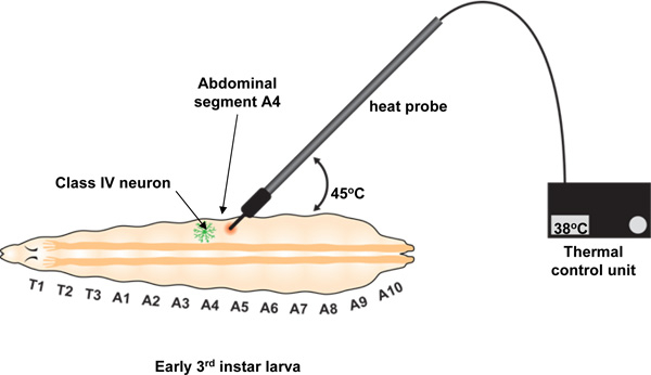
We deliver a noxious thermal stimulus to an individual larval body segment using a custom-built thermal probe manufactured by Pro-Dev Engineering (see Table of materials). Although this probe has optimal design features (a small metal tip of ~ .07 mm2 area and the ability to precisely maintain a set point temperature from 23 °C to 65 °C) in principle any tool with a small tip that can be heated to a defined temperature for a period of up to 20 seconds should suffice. The probe tip is used to stimulate early 3rd instar larvae precisely on the dorsal midline at abdominal segment A4 (see Figure 1). In response to this thermal stimulus, larvae will generally exhibit an aversive withdrawal behavior of rolling laterally by 360 degrees or more. This behavior is distinct from their light touch response to a non-noxious room temperature metal probe which generally involves a brief pause in their locomotory activity.
Protocol for the heat probe assay:
- Pre-set temperature of the thermal probe to desired set point.
- Use a paintbrush or forceps to gently transfer an individual larva onto a flat platform (we usually use a small piece of vinyl cut from a binder) on which the larvae will be subsequently stimulated. The larva should be covered by a thin film of water before contact with the thermal probe. The film of water covering the larva should be as little as possible and completely cover the larva, while ensuring that the larva is not dry when touching the vinyl.
- Gently press the probe tip against the larva at segment A4 applying light pressure with the tip at about a 45° angle between the probe and the surface of the larva (see Figure 1). The pressure should cause a slight indentation on the surface of the larva and will usually be sufficient to prevent locomotion. If the larva continues to move, apply slightly more pressure and it will usually stop. Do not record data from larvae that move beyond the field of view or for which constant probe contact cannot be achieved until response or the 20 s cutoff.
- Continue stimulating the larva until a withdrawal response is exhibited or until the 20 second cutoff is reached, whichever occurs first. Responding larvae typically first show a preliminary behavior of lifting the head and tail. This is usually followed by the withdrawal behavior of rolling at least 360 degrees. Only a complete roll of 360 degrees is scored as nocifensive behavior (the preliminary head or tail raise is not).
- Once the withdrawal behavior is initiated release contact with the probe and record the latency or time to withdrawal. If no withdrawal behavior is observed within 20 seconds, then the larva is a non-responder. The responders can be further divided into 2 categories. If the withdrawal behavior is shown within 5 seconds, then the larva is a fast-responder. If the withdrawal behavior is shown between 5-20 seconds, then the larva is a slow-responder (see Figure 1 in Babcock et al., Curr. Biol. (2009)).